Powered by Blogger.
RSS

Welcome

I created this blog as an instrument of what I have encountered in the world of veterinary medicine as a proud vet student. Comments and suggestions are welcome here at;

sweet_daffodil90@yahoo.co.uk

Regards,
Aina Meducci 2012

Disclaimer

The following blog posts is not genuinely from my research but through readings and citation from trusted website. I do not own any of the copyright and therefore you may use it at your own risk

SINCE I AM NOT A VETERINARIAN YET, THEREFORE I CAN'T CONSULT ANY MEDICAL ADVICE TO YOU AND YOUR PETS! EXTREMELY IMPORTANT!.

Happy reading!
Showing posts with label dog. Show all posts
Post Icon

Horner's syndrome

I dropped into these words at last semester during my ophthalmology class. But it is then repeated in our neurological class. Seems to have interrelation on it right?

**********************************************************


Horner's syndrome in a cat

Horner's Syndrome is a common neurological disorder of the eye and facial muscles, caused by dysfunction of the sympathetic nervous system. The condition usually occurs suddenly and without warning. The most common clinical signs of Horner's Syndrome are
  • Drooping of the eyelid on the affected side (ptosis)
  • The pupil of the affected eye will be constricted (miosis), or smaller than usual
  • The affected eye often appears sunken (enophthalmos)
  • The third eyelid of the affected eye may appear red and raised or protruded (conjunctival hyperemia)

horners_syndrome_in_cats1


Horner's syndrome is not a disease but a CONDITION!


What causes Horner's syndrome?

If the sympathetic nervous system that supplies the eyes is damaged or is malfunctioning, the parasympathetic system 'takes over', and the symptoms of Horner's Syndrome appear. The damage or malfunction can occur in the neck area, the ear area or the eye area. Damage may occur because of blunt force trauma such as that caused by an automobile accident or because of a bite wound from another animal.

Other problems that can cause damage or inflammation of the nerves in this area include diseases within the eye or in the retrobulbar (behind the eye) area, problems in the middle ear (otitis media), or tumors in the chest, neck or brain.In dogs, Horner's Syndrome is 'idiopathic', which means it has an unknown cause, in about half the cases. In cats, however, a cause is virtually always found, and idiopathic Horner's Syndrome is very rare. With many cats that develop Horner's Syndrome there is a recent history of trauma, particularly being hit by a car.

Nasopharyngeal polys removal in cats can also cause the development of Horner Syndrome due to nerve damage. It is usually temporary and does not effect cat's behavior.

Depending on the cat's recent history and other physical findings on examination, veterinarian may recommend a series of diagnostic tests to determine if there is an underlying cause. Initial tests will usually include a neurologic evaluation, an otoscopic examination (examination of the ears) and x-rays of the chest and neck area.

How can sympathetic damage occurs?


Chart showing Horner's Syndrome


The nerve carrying the tiny nerve fibers that provide sympathetic control to the eye have a long path. The damage may have occurred anywhere along this path.The nerves originate in the brain stem and travel down the spinal cord in the neck area exiting just inside the chest (at the level of the second thoracic vertebra). The nerves then form the cervical sympathetic trunk, a bundle of nerves that travels back up the neck, this time outside the spinal cord, to the middle ear (this area is shown in red).These nerves then connect to new nerves just below the ear. The new nerves continue their journey to the eye. The damage can occur in the neck area, the ear area or the eye area. Damage can occur in the form of trauma, tumor involvement, infarction (abnormal blood clot), middle ear infection, or diseases of the eye itself.


Diagnosis

Localizing which area of the sympathetic nervous system is affected goes a long way in determining the nature of the damage as different areas of the system are prone to different types of injury. Certain eye drops can be used to stimulate different areas of the nervous system and determine if the lesion is in the first nerve segment or in the second nerve segment. Most injuries turn out to be in the second nerve segment.

With second nerve segment involvement: If ear infection is not obvious and disease of the eye beyond the Horner's syndrome itself is not obvious, then it is probably prudent to allow the syndrome to resolve on its own. This usually occurs within 6 to 8 weeks. Further diagnostics may be undertaken if new developments occur or if the syndrome persists beyond this time.

With first nerve segment involvement: Involvement of the first nerve segment indicates a problem in the chest or spinal cord and is more significant. Chest radiographs should be taken to rule out cancer spread to the chest (the only sign of this may be the Horner's syndrome). The front leg should be carefully checked for evidence of function loss as a tumor or protruding intervertebral disc could be exerting pressure on the spinal cord. Trauma to the neck as with a strong jerk from a collar or straining against a leash can also produce Horner's syndrome from this section of the nerve.

Generally more diagnostic work is needed for cases involving the first nerve segment as there is potential for more serious underlying causes. If the syndrome stemmed from pulling on the leash, it should resolve uneventfully, depending on how badly damaged the nerve is.


Treatment

Most cases of Horner’s Syndrome will resolve spontaneously. It is important to treat any underlying disease. There are several diagnostic tests that will be performed to determine if there is an underlying cause in your pet. Symptomatic treatment usually involves phenylephrine drops placed in each eye every 12-24 hours to dilate the pupil. Other eye medications might be useful to avoid development of corneal ulcers from the exposed keratitis.

horners_syndrome_in_cats_2


Prognosis and recovery rate

The prognosis depends on the underlying cause. Patients with chest trauma tend to have a quicker recovery rate (days to weeks) than patients with other lesions. If the underlying problem is Feline Dysautonomia, the prognosis is poor.


Sources: The pet health library; Wendy C brooks, Horner's syndrome in cat VCA Animal hospitals, Nasopharyngeal polyps in cats; ACVS Veterinary surgeon Karen Tobias

  • Digg
  • Del.icio.us
  • StumbleUpon
  • Reddit
  • RSS
Post Icon

Cardiopulmonary resuscitation (CPR) in animal

Animal CPR (a big LOL)

During anesthetic class, we were taught to perform CPR on emergency anesthesia. This will not happen if the anesthetists are very careful in monitoring the patient throughout surgical period. So, let's dive in!

**********************************************************


CPR in dog


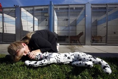
CPR is an emergency technique used to help someone whose heart and/or breathing has stopped. Although somewhat modified, the same techniques used for people – rescue breathing and chest compression – can be used to help treat an animal in distress.

The first lesson to know about CPR is that it doesn't restart a stopped heart. The purpose of CPR, in both humans and animals, is to keep them alive until the heart begins beating on its own or a cardiac defibrillator can be used. In animals, CPR is frequently unsuccessful, even if performed by a trained veterinarian. Even so, attempting CPR will give animal fighting chance.


PCR on cat


At first, we must know the ABC rules!


A-Airway
1.Listen and feel to determine if your pet is breathing/has a pulse.
2.Carefully pull the tongue straight out of the animal’s mouth to open the airway
3. Make sure that the neck is reasonably straight; try to bring the head in-line withthe neck.

WARNING: Do not over-straighten the neck in cases where neck/head trauma exists

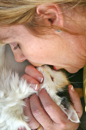
4. Attempt 2 rescue breaths, by closing the mouth, and performing mouth-to-nose ventilations. If they go in with no problems continue to B-Breathing.

Step 2: Place pet on his right side (legs away from you)...
Opening airway in dog

See above for positioning to give breaths to a cat.

Opening airway in cat

5. If not, reposition the neck and try5. step 3 again.

6.Visibly inspect the airway by looking into the mouth, and down the throat for foreign objects occluding the airway. Unlike human-CPR, rescuers may reach into the airway and remove foreign objects that are visible.

6. If you still can’t breath into the animal,proceed to the Heimlich maneuver.

Heimlich maneuver

Use to remove choking material that cause inability in breathing

1. Turn the animal upside down, with its back against your chest 2. Hug the animal with your fist in your hand, just below the rib-cage ( for cats, just squeeze 1 hand in the same place)


2. With both arms, give 5 sharp thrusts (bear hugs) to the abdomen. Perform each thrust as if it is the one that will expel the object

3. Stop, check to see if the object is visible in the airway, if so, remove it and give 2 mouth-nose rescue breaths. If the breaths do not go in, go back to step 1


Heimlich maneuver


B-Breathing

After achieving a patent airway, one must determine whether the animal is breathing, and whether this breathing is effective:

1. Carefully pull the tongue straight out of the animal’s mouth to open the airway

2. Make sure that the neck is reasonably straight; try to bring the head in-line withthe neck.

3. Breathe at 12 breaths per minute (1 every 5 seconds)With each breath just make the chest rise (do not overinflate, expecially on a small animal)

IMPORTANT: If the breaths do not go in, stop and return to A-Airway!


Proceed to C-Circulation, while continuing breathing support as necessary.


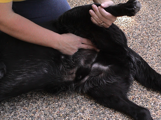
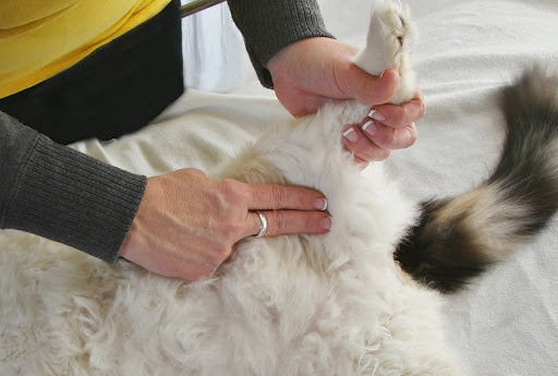
Check the femoral pulse!

Step 3 (Dog): Check pulse at femoral artery (where hind leg meets torso), If no pulse…

Dog

Step 3 (Cat): Check pulse at femoral artery (where hind leg meets torso), if no pulse…

Cat


C- Circulation

This is the final step of CPR and should only be started after the A-airway and B-breathing steps have been completed:

1. Make sure that there are no major (pooling/spurting blood) points of bleeding. Control as necessary by applying pressure with your hand

2. Check for a pulse in the groin (check carefully on a conscious dog or cat!)

3. Lay the animal on its right side

Step 4 Gently take animal’s left front leg and bend it at the elbow, rotating it at the shoulder.  Where his elbow touches his body is where you place your left hand for compressions.


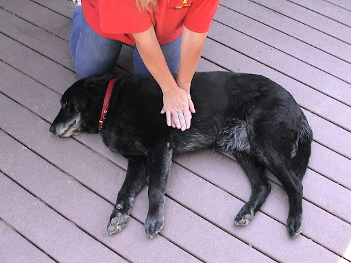
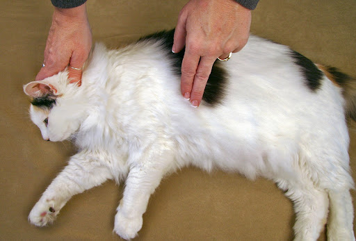
4. Locate your hands where its left elbow touches the chest, approximately the middle of the rib-cage (for cats use 1 hand in asqueezing motion).

Compressions for medium to large dogs: With you left hand, push on the chest 10-15 times (approximately 3 compressions every 2 seconds) and then deliver 2 more breaths. Repeat.  Every 4 cycles, check for a pulse.


Location of heart

Compress the chest 15 times followed by 2 rescue breaths (3 compression every 2 seconds). Deliver 2 more breath. Repeat. Every 4 cycle for a pulse.

Compressions for small cats and dogs:  Use fingertips to compress heart in place of your left hand or place 4 fingers of your left hand under the animal’s chest and compress on the top with left thumb: 5 compressions/1 breath and check for a pulse every 8-10 cycles.

Compression for small cats and dogs: Use fingertips to compress heart in place of your left hand or place 4 fingers of your left hand under the animal’s chest and compress on the top with left thumb: 5 compression/1 breath and check for a pulse every 8-10 cycles.

If there are two people, one breathes and the others compresses at the rate of one breath for every 2-3 compression


After the CPR is successfully performed, quickly send the animal to the nearest vet!


Precaution in CPR

1. Take care not to over inflate the lungs, observe that you ventilate with a normal rise and fall to the chest.

2. Never breathe or compress on an animal that is breathing or has a pulse.

3. When doing compression, realize you must compress 1/4 to 1/3 the width of the chest. You must flex the ribs, press the lungs in order to squeeze the blood out of the heart and release so that it can flow around and back into the other chamber.

4. For barrel-chested breeds, you may position dog on his back and compress chest human-style (hand on top of hand over chest): 15 compressions/2 breaths checking for a pulse every 4 cycles.

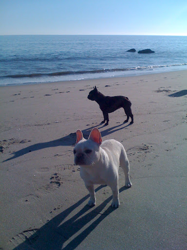
For barrel-chested breeds (like Fala and Winnie above) you may position dog on his back and compress chest human-style (hand on top of hand over chest):  15 compressions/2 breaths checking for a pulse every 4 cycles.

Barrel-chested dog


**Perform CPR until you have reached a veterinary hospital. After 20 minutes, however, the chances of reviving an animal are extremely unlikely.


Maybe you can learn something here..


Sources: Sheraton luxuries; CPR guide, CPR in dogs;petplace.com, dailywag; Martha steward.com; Pet first aid

  • Digg
  • Del.icio.us
  • StumbleUpon
  • Reddit
  • RSS