Powered by Blogger.
RSS

Welcome

I created this blog as an instrument of what I have encountered in the world of veterinary medicine as a proud vet student. Comments and suggestions are welcome here at;

sweet_daffodil90@yahoo.co.uk

Regards,
Aina Meducci 2012

Disclaimer

The following blog posts is not genuinely from my research but through readings and citation from trusted website. I do not own any of the copyright and therefore you may use it at your own risk

SINCE I AM NOT A VETERINARIAN YET, THEREFORE I CAN'T CONSULT ANY MEDICAL ADVICE TO YOU AND YOUR PETS! EXTREMELY IMPORTANT!.

Happy reading!
Post Icon

Runting and Stunting syndrome

I'm going to broiler farm for avian trip. Here's the chief complaint.

****************************************************************


Same age but different growth stage


Runting and stunting syndrome

Few things are well defined regarding runting-stunting syndrome.  Starting by its  very name, “runting stunting” is poorly defined and will probably be changed in the future depending on the etiological agent o agents identified.  In poultry, a “runt” is defined as an animal that is unusually small, especially the smallest of a flock.  “Stunting” relates to the hindering of the normal growth of an individual.  Thus, “runting-stunting” can be defined as a syndrome in which a number of individuals in a flock appear considerably small due to delayed growth.  The larger the number of chickens that are unusually small, the lower the body weight average for the flock.


Runting-stunting syndrome (RSS) has been recognized for many years and was first described in the 1970s.  One can think of a seemingly endless list of possible causes for RSS, including faulty poultry genetics, management, environmental challenge, feeding and nutrition, infectious disease and perhaps other reasons and any possible combination of them.  


Although some features of RSS have been reproduced upon experimental infection with reovirus, the general consensus is that these viruses are unlikely to be the sole culprit in RSS.  Small round viruses (small within the context of general virology) have been visualized many times in the intestines and intestinal contents of affected broiler chickens using electron microscopy, but such viruses can also be found in nonaffected flocks or individual chickens.  

This condition is usually seen between 3-6 weeks of age and is usually observed in meat type chickens.


Brooding at cool temperatures tends to worsen RSS symptoms, as does short down-time between flocks. Certain strains of birds appear to be more susceptible to the effects of RSS than others and male birds are more severely affected than females (Zavala and Barbosa, 2006). However, it is interesting to note that researchers have found that resistant broiler strains have stronger immunological responses than susceptible strains. This difference is particularly pronounced when gut immunity is compared (Rebel et al., 2006). Some researchers have suggested that the poor growth and retarded feathering (which are consistently observed in RSS cases) are due to a common underlying infection, while virtually all other symptoms result from other infections or management factors.


Clinically, affected flocks show large numbers of immobile chicks huddling around the feeders and drinkers within hours after placement.  Some may peck incessantly at the walls.  The litter quickly becomes damp and chicks may exhibit matted down in the abdominal area as a result of resting on wet litter.  Consumption of chick starter feed typically lasts a day or two longer than usual.  As early as 6-7 days of age many chicks will already appear stunted, pale and sometimes disoriented, but the usual peak of the problem occurs at around 10-12 days of age.  The bodies of affected chicks will look small relative to the length of the primary feathers of the wings and beak.  A small number of affected chicks may display “helicopter” feathers in their wings and other feather abnormalities.   


“Helicopter” wing feathers


Some of the internal gross lesions or changes that have characterized recent cases of RSS include (not exclusively):  small livers with a grossly enlarged gall bladder; pale, thin, almost translucent intestinal wall with wet pellets of undigested food that is visible through the intestinal wall; large amount of fluids inside the small and large intestines; infrequent feed impaction in the cloaca; occasional increased amount of pericardial fluid;  sporadic white or cream-colored plaques in individual proventricular glands. 


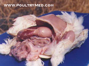
Proventriculitis or proventricular distension. The poventriculus is large almost as the gizzard.


Click here to ZOOMRunting and Stunting Syndrome

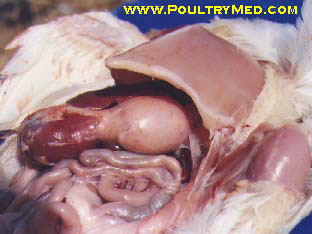
Diffuse enlargement of the poventriculus due to thickening of its wall. 


Click here to ZOOMRunting and Stunting Syndrome

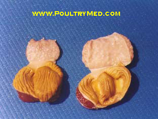
Normal (left) and affected (right) poventiculus. 


Various microscopic lesions have been described but the most frequent one is the presence of multiple cysts involving the intestinal crypts.  The early lesion can described as “cystic enteropathy” and as it evolves into an inflammatory lesion it turns into “cystic enteritis”.  The nature of the inflammatory responses suggests a viral etiology rather than bacterial.  As the lesion progresses it may result in shortening and clubbing of the intestinal villi.  Other microscopic lesions are important observations but they are not always present in cases of RSS.  Viruses, nutritional deficiencies and parasites have been proposed as possible etiologies of cystic enteropathy.  Until now, only deficiencies of some complex B vitamins (thiamin, riboflavin, pantothenic acid, and niacin) have consistently reproduced cystic enteropathy.  


Treament

No treatment available


Control

When RSS is reported in an area, it is important for the industry in the area to tighten Biosecurity procedures to reduce the possibility of exposure and to slow the spread of the disease. It is particularly important to emphasize procedures that control farm visitors, properly manage disposal of mortality and limit vermin infestations (rodents, wild birds and insects). 

The objective of proper poultry house management is to provide an environment for the birds that is virtually stress free. In RSS situations, poultry house management is doubly important. Good management starts before the birds arrive. A minimum of 12 days of downtime should be allowed between flocks. Since litter has been shown to transmit the disease, it should be removed if birds have broken with RSS. If it is not possible to remove the litter, heat the litter to 100°F for 100 hours or compost the litter in the poultry house to lessen the possibility of passing the disease to the next flock via litter. The brood chamber should be cleaned and disinfected as thoroughly as possible prior to chick placement. Since low brooding temperatures have been shown to worsen the effects of RSS, DO NOT reduce brooding temperatures to save fuel. Check on birds often and maintain a house environment that is as stress free as possible. Remove dead birds quickly and cull severely if RSS breaks. The application of vinegar or other acidifiers via water may reduce spread of the disease. Supplemental vitamins and minerals in both breeder and broiler feeds has also been shown to improve immunity in chicks and their ability to deal with RSS. 

Certain strains of reovirus (e.g. 1733 and 2408) were originally implicated as the cause of RSS and vaccines have been developed for such strains. While vaccination of broilers for RSS may be effective about 50% of the time, a consistent vaccination program for breeders often provides long term benefits (Shane, 2008, van der Heide, 2000). RSS vaccination programs for breeders generally provide protection for adult birds, reducing the possibility of spread to young birds. In addition, immunity in breeder hens is passed to chicks, helping to protect them from the disease


Sources

The poultry informed professional; www.poultry-health.com, Runting and stunting syndromes in broilers; www.poultrysite.com, REO Virus (Runting and stunting syndrome)www.vetsoft.com, Runting ans stunting syndrome; www.poltrymed.com


  • Digg
  • Del.icio.us
  • StumbleUpon
  • Reddit
  • RSS
Post Icon

Avian disease: Infectious coryza

Recently I have encountered an avian case of which we cant really tell what it is cos we're still investigating the case. But from our differential, infectious coryza is one of them. The chicken was found with swollen face plus respiratory problems.


*************************************************************************


Swollen face


Infectious coryza
A usually acute, sometimes chronic, highly infectious disease of chickens, occasionally pheasants and guinea-fowl, characterised by catarrhal inflammation of the upper respiratory tract, especially nasal and sinus mucosae.


Aetiology

The causative bacterium, Haemophilus paragallinarum (gallinarum) is a gram-negative, pleomorphic, nonmotile, catalase-negative, microaerophilic rod that requires nicotinamide adenine dinucleotide (V-factor) for in vitro growth. When grown on blood agar with a staphylococcal nurse colony that excretes the V-factor, the satellite colonies appear as dewdrops, growing adjacent to the nurse colony.


Chocolate agar is an excellent Haemophilus growth medium as it allows for increased accessibility to these factors.Alternatively, Haemophilus is sometimes cultured using the "Staph streak" technique: both Staphylococcus and Haemophilus organisms are cultured together on a single blood agar plate. In this case, Haemophilus colonies will frequently grow in small "satellite" colonies around the larger Staphylococcus colonies because the metabolism of Staphylococcus produces the necessary blood factor by-products required for Haemophilus growth.


Hemophilus on blood agar plate


Transmission

Chronically ill or healthy carrier birds are the reservoir of infection. Chickens of all ages are susceptible, but susceptibility increases with age. The incubation period is 1-3 days, and the disease duration is usually 2-3 wk. Under field conditions, the duration may be longer in the presence of concurrent diseases, eg, mycoplasmosis.

Infected flocks are a constant threat to uninfected flocks. Transmission is by direct contact, airborne droplets, and contamination of drinking water. Commercial farms that have multiple-age flocks tend to perpetuate the disease. Egg transmission does not occur (no vertical transmission).

The bacterium survives 2-3 days outside the bird but is easily killed by heat, drying and disinfectants. Intercurrent respiratory viral and bacterial infections are predisposing factors.


Clinical signs

Facial swelling.
Purulent ocular and nasal discharge.
Swollen wattles.
Sneezing.
Dyspnoea.
Loss in condition.
Drop in egg production of 10-40%.
Inappetance


Infectious Coryza

Facial swelling: Common finding


Purulent discharge. Note the solid white material under the eye

Preview Image

Swelling of infraorbital sinuses and eye discharges


Post-mortem lesions
  • Catarrhal inflammation of nasal passages and sinuses.
  • Conjunctivitis.
  • Eye-lid adherence.
  • Caseous material in conjunctiva/sinus.
  • Tracheitis.

Preview Image

Severe fibrinosuppurative conjunctivitis


Preview Image


Infraorbital sinus: Catarrhal and suppurative sinusitis


Preview Image


Air sacs: Severe diffuse caseous airsacculitis (uncommon finding)


The histopathologic response of respiratory organs consists of disintegration and hyperplasia of mucosal and glandular epithelia and edema with infiltration of heterophils, macrophages, and mast cells.


Differential diagnosis

  • Mycoplasmosis
  • Infectious Laryngotracheitis
  • Newcastle disease
  • Infectious bronchitis
  • Avian Influenza
  • Swollen head syndrome
  • Vitamin A dificiency

Diagnosis

Isolation of a gram-negative, satellitic, catalase-negative organism from chickens in a flock with a history of a rapidly spreading coryza is diagnostic. The catalase test is essential, as nonpathogenic Haemophilus organisms, which are catalase-positive, are present in both healthy and diseased chickens. A PCR test that can be used on the live chicken and that has proved superior to culture, even in developing countries, has been developed. Production of typical signs after inoculation with nasal exudate from infected into susceptible chickens is also reliable diagnostically. No suitable serologic test exists; a hemagglutination-inhibition test is the best of the available tests.


Treatment

Erythromycin and oxytetracycline are usually beneficial. Several new-generation antibiotics (eg, fluoroquinolones, macrolides) are active against infectious coryza. Various sulfonamides, sulfonamide-trimethoprim, and other combinations have been successful but must not be used in layers. In more severe outbreaks, although treatment may result in improvement, the disease may recur when medication is discontinued.

Preventive medication may be combined with a vaccination program, if started pullets are to be reared or housed on infected premises.


Control

Prevention is the only sound method of control. “All-in/all-out” farm programs with sound management and isolation methods are the best way to avoid the disease. Replacements should be raised on the same farm or obtained from clean flocks. If replacement pullets are to be placed on a farm that has a history of infectious coryza, bacterins are available to help prevent and control the disease.


Sources:

The merck's veterinary manual, Altlas of Avian diseases, Cornell university, college of veterinary medicine. Infectious coryza;www.poultrysite.com.


  • Digg
  • Del.icio.us
  • StumbleUpon
  • Reddit
  • RSS
Post Icon

Hemostasis: Blood clotting mechanism

I'm not feeling good . We had a brief discussion with professor regarding pathogenesis of anthrax, the toxin production, ect ect ect. It was pretty good at first because we are discussing but it turned us down because he asked about BLOOD CLOTTING MECHANISM. Oh my god, I hardly remember any of them :( It made me realised that there are many things i have left out. Oh my,,


*********************************************************


How does the bleeding is stopped?


Blood coagulation refers to the process of forming a clot to stop bleeding. There are 3 mechanisms that work together to stop the flow of blood. They are;

1. Vasoconstriction
2. Platelet plug formation
3. Clotting of blood


Vasoconstriction of a damaged blood vessel slows the flow of blood and thus helps to limit blood loss. This process is mediated by:

Local controls. Vasoconstrictors such as thromboxane are released at the site of the injury.
Systemic control. Epinephrine released by the adrenal glands stimulates general vasoconstriction

 Platelet plug. Platelets aggregate to the site of the injury. They stick together acting as a "plug." Platelets also activate the process which causes a fibrin clot to form, known as secondary hemostasis. Platelets stick to collagen and become activated. Activated platelets release chemicals such as ADP, and thromboxane, that cause the aggregation of more platelets to the site of injury. Platelet aggregation results in the formation of a platelet plug which acts to stem the flow of blood from the broken vessel. It is essential that platelets become activated only at the site of a broken vessel. Otherwise activated platelets would form plugs and induce clots in inapropriate places. Healthy vessels secrete an enzyme called prostacyclin that functions to inhibit platelet activation and aggregation.


Secondary hemostasis

Platelets alone are not enough to secure the damage in the vessel wall. A clot must form at the site of injury. The formation of a clot depends upon several substances called clotting factors. These factors are designated by roman numerals I through XIII. These factors activate each other in what as known as the clotting cascade. The end result of this cascade is that fibrinogen, a soluble plasma protein, is cleaved into fibrin, a nonsoluble plasma protein. The fibrin proteins stick together forming a clot. 


The blood contains about a dozen clotting factors. These factors are proteins that exist in the blood in an inactive state, but can be called into action when tissues or blood vessels are damaged. The activation of clotting factors occurs in a sequential manner. The first factor in the sequence activates the second factor, which activates the third factor and so on. This series of reactions is called the clotting cascade.


Blood clotting is the transformation of liquid blood into a semisolid gel. Clots are made from fibers (polymers) of a protein called fibrin. Fibrin monomers come from an inactive precursor called fibrinogen. The body of the fibrinogen molecule has caps on its ends that mask fibrin-to-fibrin binding sites. If the caps are removed then fibrin monomers polymerize to form fibrin polymers. This process requires thrombin, the enzyme that converts fibrinogen to fibrin. This process also requires calcium, which acts as a kind of glue to hold the fibrin monomers to each other to form the polymeric fiber. The fibrin fibers form a loose meshwork that is stabilized by clotting factor XIII. The stabilized meshwork of fibrin fibers traps erythrocytes, thus forming a clot that stops the flow of blood.


The clotting cascade occurs through two separate pathways that interact, the intrinsic and the extrinsic pathway. 
 

Extrinsic Pathway

The extrinsic pathway is activated by external trauma that causes blood to escape from the vascular system. This pathway is quicker than the intrinsic pathway. It involves factor VII.

Intrinsic Pathway

The intrinsic pathway is activated by trauma inside the vascular system, and is activated by platelets, exposed endothelium, chemicals, or collagen. This pathway is slower than the extrinsic pathway, but more important. It involves factors XII, XI, IX, VIII. Damage to the vessel wall stimulates the activation of a cascade of clotting factors (for the sake of simplicity we will not consider the individual factors). This cascade results in the activation of factor X. Activated factor X is an enzyme that converts prothrombin to thrombin. Thrombin converts fibrinogen to fibrin monomers, which then polymerize in fibrin fibers. Fibrin fibers form a loose meshwork that is stabilized by crosslinks created by factor XIII. The stabilzed meshwork of fibrin fibers is now a clot that traps red blood cells and platelets and thus stops the flow of blood.

Common Pathway

Both pathways meet and finish the pathway of clot production in what is known as the common pathway. The common pathway involves factors I, II, V, and X.


Summary of blood clotting mechanism


Inhibition of blood clotting

It would be dangerous if blood clotting were to continue to expand beyond the boundaries of the injury. Excessive clotting is inhibited because:
Clotting factors are rapidly inactivated. There are enzymes in the blood that function to inactivate clotting factors. As clotting factors are taken away from the site of injury by the blood stream, they become inactivated by these enzymes. This ensures that clotting will only occur at the site of injury and not progress steadily down the vessel.
Fibrin fibers inhibit the activity of thrombin. Thrombin acts to convert fibrinogen to fibrin, yet fibrin fibers have an inhibitory effect on the activity of thrombin. As the clot grows this inhibition intensifies. This constitutes a negative feedback loop, where the product of thrombin activity (fibrin) feeds back to shut off thrombin.


Clot Removal

Blood clots are designed to be temporary. After the clot has formed, the process of vessel repair begins. Epithelial cells at the margin on the injury undergo cell division. These new cells eventually fill the gap in the vessel created by the injury. Also, cells called fibroblasts are recruited to the area. Fibroblasts form connective tissue that repairs the basement membrane of the vessel (fibroblasts also form scar tissue that may or may not be removed over time). At this point the vessel is healed and the blood clot is no longer needed. 


The clot itself stimulates the secretion of tissue plasminogen activator (TPA) from the surrounding vascular epithelium. TPA is an enzyme that catalyzes the conversion ofplasminogen to plasmin. Plasminogen is an inactive precursor molecule found in the blood, but plasmin is an enzyme that dissolves clots. Plasmin levels are not very high so clot removal is a slow process. By the time the clot has been completely dissolved by plasmin, the vessel has had a chance to heal itself. In summary, the clot, which forms rapidly, calls for its own destruction by initiating the activation of plasmin.


Anticoagulants are substances that inhibit the process of clotting.

  1. Heparin
    • produced primarily in the liver and lung (usually obtained from pigs or cows)
    • acts as an anticoagulant by inhibiting the activity of thrombin
    • Used clinically for acute problems, also used to prevent clotting in IVs (heplock)
    • fast acting, but short-lived drug. It must be injected to be effective.
  2. Coumadin (dicoumarol, warfarin)
    • taken orally in small doses for long-term control of blood clotting
    • acts as an anticoagulant by inhibiting the processing of vitamin K, which is required for the synthesis of several clotting factors including prothrombin.
    • It is slow acting, requiring days to have an effect.
  3. Citrates
    • Calcium is required for polymerization of fibrin.
    • Citrates bind up (chelate) calcium and thus inhibit the formation of clots
    • Citrates are used in long term blood storage.
    • Transfusion of large amounts of citrate-containing blood can be dangerous. The citrates chelate calcium in the body and thus disrupt processes such as nerve transmission and muscle contraction.


Blood Clotting Disorders

Von willebrand disease
Animal with this inherited disorder bleed excessively due to an inability to rapidly form blood clots. This defect could be caused by a deficiency of any of the clotting factors.

Vitamin K deficiency
Vitamin K is essential to the maturation of several clotting factors including factor X and prothrombin. In the absence of vitamin K these clotting factors are defective and thus inhibit the clotting mechanism. 


References: Mechanism of blood clotting;http://departments.weber.edu/chpweb/hemophilia/mechanisms_of_blood_coagulation.htm, Homeostasis; http://www.biosbcc.net/doohan/sample/htm/Hemostasis.htm


  • Digg
  • Del.icio.us
  • StumbleUpon
  • Reddit
  • RSS
Post Icon

The Modified Robert Jones Bandage

In class, we were taught to demonstrate modified Robert Jones Bandage with a cat. This is just a quick revision before I started my clinical industrial training shortly.


Bandaging a cat using MRJB


The Robert Jones bandage is a common external splint applied to a limb for the temporary support of a fracture before surgical intervention can occur. It is used to treat many canine and feline limb injuries (e.g., tibial fractures, severe limb lacerations). It promotes healing by immobilizing the injured area, thereby limiting swelling and providing protection from secondary trauma. Compared with other padded bandages, the Robert-Jones bandage offers limb stability, tissue fluid absorption, and protection from trauma. Generally, most of the compression is lost after several hours to days as the cotton loosens. For definitive support of a fracture the bandage should never be on longer than a few days without reinforcement using rigid splint material.

The Modified Robert Jones bandage can be applied for the temporary compression of a limb after surgery. It functions to protect the incision site and decrease swelling to aid in healing.


Materials needed


Stirrup tape


Cotton roll (Plenty of them)


Surgical cast padding


A Vet wrap (only 1)


Let's just says a dog with fracture hindlimb underwent surgery to correct the bone. This is the steps on how the animal should be bandage to protect the bone. The 5 very simple step.


Step 1

Step 1: Using white bandage tape, place stirrups on the distal 1/3rd of the limb overlapping the toes and extending approximately an equal length from the end of the leg. Be sure to tab the ends for easy separation later on.


Step 2

Step 2: Wrap the leg lightly with cast padding starting at the toes and moving proximally. Overlap the bandage 50% as you wrap and try to get 2 layers of padding. Note - it is important not to exceed approximately two layers of cast padding. Excessive padding will cause premature loosening of the bandage as the cotton compresses overtime.


Step 3


Step 3: Wrap the leg tightly with a conforming bandage starting at the toes and moving proximally. This is the step where you create compression, however, not as much as you would with the standard Robert Jones bandage. Overlap the bandage 50% as you wrap and make sure the toes are still visible.


Step 4

Step 4: Separate the tape stirrups, rotate them proximally, and secure them to the compression bandage thus creating a barrier and preventing the bandage from slipping down. Note - you should always make sure some of the underlying cast padding is visible on the end by the toes.


Step 5

Step 5: Wrap the leg in vet wrap starting at the toes at an angle to cover the distal ends of the bandage and again moving proximally as you progress. Be sure to overlap 50% as you apply the material.


Sources: Directly taken from:
http://cal.vet.upenn.edu/projects/orthopod/csfr/terms/modifiedrobertjonesbandagecipage.htm


  • Digg
  • Del.icio.us
  • StumbleUpon
  • Reddit
  • RSS
Post Icon

Pyrethrins and pyrethroids toxicity

Just about going to sleep when suddenly my brother says our persian-mixed cat licking the excess chrysanthemum tea from his cup. I told him no! because I remembered what our prof told us in class regarding chrysanthemum toxicity. See, being a vet makes you know everything about do and dont's in animal!


**************************************************************


Nice tea..but,,


What are Pyrethrins?

Extracted from dried flowers of the chrysanthemum cinerariaefolium, natural pyrethrins are insect repellents. Both natural pyrethrins and synthetic ones called permethrins are used in insecticides and pet flea and tick products.


Chrysanthemum cinerariaefolium


What are pyrethroids?

Pyrethroids (allethrin, alphacypermethrin, bioremethrin, cypermethrin, deltamethrin, fenvalerate. permethrin, phenothrin, resmethrin, tetramethrin) are synthetic pyrethrins.

Pyrethrins and pyrethroids are used in as insecticides for the treatment of endoparasitic infestation. They are use in cats, dogs, sheep, cattle, poultry, horses, goats and humans. They are used in dogs for the control of fleas and lice. Preparations are available as dusting flowers, shampooa and sprays.


Flea spray containing pyrethrin


Shampoo containing pyrethrin


Toxic Dose

Varies depending upon type of pyrethroid, size of animal, and species.


Mechanism of toxicity

Toxicity may arise from self-grooming of treated hair. Occasionally, toxicity may arise from animals being in close association with or grooming treated animals


The toxic effects of pyrethroids and pyrethrins are due to alteration of the kinetics of voltage dependent sodium channels in nerves membrane, which causes repetitive discharge or membrane depolarisation (sodium unable to get out). Some pyrethroids may also inhibit GABA receptors causing loss inhibition. This can lead to hyperexcitability of nervous tissues and maybe the mechanism by which these compound produce convulsion.


Convulsion (seizures)


Natural pyrethrins are broken down by digestive juices, while the liver metabolizes synthetic pyrethrins before they enter an animal's GI tract. Pyrethrin toxicity in cats occurs because their livers are incapable of properly metabolizing synthetic pyrethrins.

All pyrethrins are easily hydrolyzed and degraded by stomach acids so toxicity following ingestion by pets is very low. Toxicities, although rare, do occur. A cat or dog with pyrethrin toxicosis generally will salivate, tremor, vomit, and may seizure. Generally, signs of toxicosis will be gone after 24 hours. Pyrethrins are some of the safest ingredients available, especially when one expects ingestion may occur, as is the case of cats and kittens. If lactating, breeding or pregnant animals must be treated for external parasites, pyrethrins are often recommended. Pyrethrins are generally safe for kittens as young as 4-6 weeks of age.


Clinical effect

Onset is usually 1-3 hours, sometimes up to 12 hours. Effects may have duration of 1-3 days. Simultaneous exposure to organophosphate insecticides may increase the toxicity of pyrethroids and pyrethrins. Piperonyl butoxide often added as a synergist to delay metabolism and increase toxicity in insects, produces effects similar to that of pyrethrins and pyrethroids. The clinical signs are shown below

  • Vomiting
  • Diarrhea
  • Ataxia
  • Tremor
  • Twitching
  • Dilated pupils
  • tachycardia
  • Pyrexia
  • Hyperexatability
  • Thirst
  • Fasciculation
  • Convulsions
  • Hyperaesthesia

Treatment

If within 2 hours of ingestion, induce vomiting or perform gastric lavage and then administer adsorbents

Emetics:

  • Xylaxine (0.2mg/kg IV, 0.5-1 mg/kg SC or IM)
  • Apomorphine (0.005 mg/kg IV/IM, 0.1 mg/kg SC)

Adsorbent
  • Activated charcoal (2 g/kg IM/SC)

Treatment thereafter is essentially symptomatic and supportive. Cats exposed to topical forms of synthetic pyrethrins should be thoroughly bathed with mild dish soap and lukewarm water. Overly warm water may enhance absorption of the pyrethrins, worsening the symptoms. For cats with minimal exposure this treatment may be enough.If the cat has absorbed lots of synthetic pyrethrins, she may need treatment with an anti-seizure medication like diazepam and/or the anti-tremor medication methocarbynol along with IV fluids to balance her electrolytes.


The body temperature is monitored, especially after bathing, as hypothermia increases the toxicity. Other treatments include anticonvulsants and/or muscle relaxants for controlling the seizures, and providing a safe environment to prevent injury resulting from the incoordination and disorientation. Atropine can be used to help decrease some of the signs such as the drooling. Fluids are generally administered.


Most pets recover from pyrethrin intoxication within 24-48 hours; recovery from pyrethroids may take longer. If no improvement is seen within 24 hours with treatment, the pet should be reevaluated.


" Pyrethrin is NOT the same as permethrin. Permethrin is a synthetic pyrethrin, and is less easily broken down than pyrethrin. Although its toxicity is relatively low, it is higher than that of pyrethrin. Pyrethrins can be used on cats; permethrins should NOT be used on cats. "


Sources: Handbook poisoning of dogs and cats, Blackwell science, pyrethrins and pyrethroid www.petseducation.com, The use of pyrethrins and pyrethroids in cats and dogs www.peteducation.com, Pyrethrins toxicity in cats.

  • Digg
  • Del.icio.us
  • StumbleUpon
  • Reddit
  • RSS
Post Icon

Why red blood cell has no nucleus?

I came across this terminologies while reading;


" Why red blood cell has no nucleus? "


Here's the answer:

While in the bone marrow, the developing red blood cells are nuclear. They include in order of maturity, the prorubricyte, rubricyte and metarubricyte (being the most matured). Just before being released from the bone marrow, the metarubicyte discard their nuclei.Therefore rbc in most domestic animal (except birds) are anuclear, biconcave, discoid cells.

Hence, with no nucleus, the number oxygen's molecule that can bind to the haemoglobin can be increased up to 4 molecules for each red blood cells.


Erythrocyte maturation diagram


Sources: Veterinary Medical Terminologies 2nd edition, Saunders Elsevier

  • Digg
  • Del.icio.us
  • StumbleUpon
  • Reddit
  • RSS
Post Icon

Infectious Canine Hepatitis

I remember during my 2nd year where we had virology oral exam with Dr J. So before waiting our names to be called in, we were scampering for notes and memorized the all the virus and the diseases associated with it like nobody business. Until my name was called, I went and sat in front of the examiner. I was doing quite well for the rest of the exam until he came out with the final question;


"Name the virus that causes infectious canine hepatitis?"


Opps...I blown the day :(


At the end of the semester, virology is my favorite subject


**********************************************************************

Canine infectious hepatitis

Infectious canine hepatitis an acute highly contagious viral disease of dogs which affect the liver and other organs and is characterized by high rise of temperature, diarrhoea, vomiting and convulsion. Dogs of all ages, specially puppies and foxes is affected by this disease. A carrier dog may recover, but continue to spread the virus via its urine for up to six months.Severely affected dogs will have a fever, loss of appetite, depression, diarrhoea, tonsillitis and acute abdominal pain due to an inflamed liver. Death may result within 36 hours. Corneal opacity known as "blue eye" may follow infection.


Abdominal pain in a dog with ICH


Aetilogy and transmission

ICH is caused by a nonenveloped DNA virus, canine adenovirus 1 (CAV-1), which is antigenically related only to CAV-2 (one of the causes of infectious canine tracheobronchitis). CAV-1 is resistant to lipid solvents and survives outside the host for weeks or months, but a 1-3% solution of sodium hypochlorite (household bleach) is an effective disinfectant.


Ingestion of urine, feces, or saliva of infected dogs is the main route of infection. Recovered dogs shed virus in their urine for ≥6 mo. Initial infection occurs in the tonsillar crypts and Peyer’s patches, followed by viremia and infection of endothelial cells in many tissues. Liver, kidneys, spleen, and lungs are the main target organs. Chronic kidney lesions and corneal clouding (“blue eye”) result from immune-complex reactions after recovery from acute or subclinical disease.


Even a dog dish that has been licked clean can carry the virus. The tonsils and lymph nodes are the first body parts effected. The incubation period can last 4-9 days, after which the virus enters the bloodstream.


dog eye diseases photo

Blue eye dog infectious hepatitis


Pathogenesis

The virus can remain in the kidneys of dogs that have recovered from infection for up to one year, making it all the more difficult to determine what locations might be contaminated. The particles enter the body through the nose or mouth and initially infect the tonsils. Here the virus replicates and proceeds to invade the nearby lymph nodes.This process of replication continues for 4 to 8 days, at which point the virus spreads from the lymphatic system and enters the bloodstream.

Once in the blood, CAV-1 particles infect a varieyt of other target organs – the liver, kidneys, and eyes. The viral particles themselves are cytotoxic which is why they are capable of doing so much damage. The liver is almost always the most severely affected organ, with consequences including disturbance of protein and fat manufacture, problems with bile secretion, and difficulty detoxifying drugs, chemicals, and bacteria from food. ICH is generally not fatal, but when it is, death is usually a result of massive hemorrhage, fluid leakage from a severely damaged liver, and central nervous system trauma which triggers seizure and coma.


INFECTIOUS CANINE HEPATITIS IS NOT ZOONOTIC!


Clinical signs

Signs vary from a slight fever to death. The mortality rate is highest in very young dog. The first sign is a fever of >104°F (40°C), which lasts 1-6 days and is usually biphasic (fever associated with 2 different sets of symptoms as it progress) . If the fever is of short duration, leukopenia may be the only other sign, but if it persists for >1 day, acute illness develops. Tachycardia out of proportion to the fever may occur.

On the day after the initial temperature rise, leukopenia develops and persists throughout the febrile period. The degree of leukopenia varies and seems to be correlated with the severity of illness. Signs are anorexia, thirst, conjunctivitis, serous discharge from the eyes and nose, and occasionally abdominal pain and vomiting. Intense hyperemia or petechiae of the oral mucosa, as well as enlarged tonsils, may be seen. There may be subcutaneous edema of the head, neck, and trunk.

Clotting time is directly correlated with the severity of illness. It may be difficult to control hemorrhage, which is manifest by bleeding around deciduous teeth and by spontaneous hematomas, because of underlying disseminated intravascular coagulation. Although CNS involvement is unusual, severely infected dogs may develop convulsions from forebrain damage; brain stem hemorrhages, resulting in paresis, are common.

The fatal form of the disease results in a sudden onset of severe symptoms. Bleeding from the nose and gums, enlarged abdomen due to fluid leaking from the liver, bloody diarrhea and vomit, seizures due to central nervous system association, disorientation, coma, and death may occur. Pets may die suddenly without any obvious illness. Infectious Canine Hepatitis is most severe and the mortality rate is highest in young dogs. Veterinary attention will need to be sought ASAP.


Lesions

Endothelial damage results in “paint brush” hemorrhages on the gastric serosa, lymph nodes, thymus, pancreas, and subcutaneous tissues. Hepatic cell necrosis produces a variegated color change in the liver, which may be normal in size or swollen. The gallbladder wall may be edematous and thickened; edema of the thymus may be found. Grayish white foci may be seen in the kidney cortex.


Diagnosis

Urinalysis and blood tests may be performed in an attempt to detect viral antibodies. Similarly, viral antibodies can be detected using immunofluorescent techniques. In addition, the ELISA test may be used to look for the presence of viral particles in the feces of a sick animal.


Treatment and management

There is no specific treatment for infectious canine hepatitis so treatment is aimed at managing the symptoms until the virus runs its course. Depending on the severity of illness, hospitalization and intravenous fluid therapy may be necessary. Antibiotics don't treat the virus but may be prescribed to ward off secondary bacterial infections. In severe cases, blood transfusions may be necessary

Management for ICH;

(a) Broad spectrum antibiotic -To prevent secondary bacterial infection
(b) Anti emetic - To stop vomiting
(c) Anti diarrhoea- To stop diarrhoea
(d) Anti serum may be tried
(e) Blood transfusion in case of severely infected dog. Dose is 5-8ml/lb of body weight by slow
intra venous infusion
(f) Fluid theraphy - NSS or DNS to restore fluid and electrolyte loss
(g) Anti pyretic – In case of high rise of temperature
(h)Vitamins therapy and protein hydrolysate- To restore vitality
(i) Care nursing of infected dogs


Prevention

  • Vaccination is the most recommended method of preventing Infectious Canine Hepatitis. While it seems logical to vaccinate using the CAV-1 virus, this can usually cause unwanted side effects such as the bluing of the eye and the shedding of virus. Vaccination with a very closely related virus, CAV-2, is much safer, and will help the dog build immunity against CAV-1. CAV-2 is also thought to play a part in a common condition called kennel cough, so vaccinating with CAV-2 would result in immunity to both conditions.


It is important that all puppies commence a vaccination program from 6-8 weeks of age and that all adult dogs have their boosters.

  • Unvaccinated dog should be kept away from public places, dogs outside household, or dirty food bowls that are left outside or belonging to dogs outside household. Keep an eye on the dog during walks to ensure he does not consume urine or feces.
  • Disinfection of contaminated areas with a bleach or iodine solution can kill the virus


Sources: Merks Veterinary Manual, animalhealthcareveterinary.blogspot, ICF; Cornell university college of veterinary medicine, ICF; petsmd.com, Dog eye diseases; dog-health-handbook.com .


  • Digg
  • Del.icio.us
  • StumbleUpon
  • Reddit
  • RSS