I was recently coming from the farm trip and the thing I enjoyed while working is blood sampling. Honestly, bleeding program is the most exciting activity for me; to see the blood oozing out from vein to the tube and you can't help the satisfied feeling because you have done it. However, I have only learn to do venipuncture to animals such as sheep, goat and cattle. I have not put a try on other animals (it'll be pleasure to practice) and here's some information about blood sampling technique in animals.
**********************************
Equipments
1. Needle
Needle size : The smaller the number, the bigger the lumen
Needle and needle sheath
2. Holder

Holder: To direct the blood from the needle to the vacutainer
3. Vacutainer tube
Containers containing coagulant (commonly used in veterinary medicine)
- Red top: Contain clot activator and gel for serum seperation
- Orange top: Contain thrombin (a rapid clot activator) for STAT serum testing
Containers containing anticoagulant (Commonly used in veterinary medicine)
- Green top: Contain sodium heparin or lithium heparin for plasma determination
- Purple top: Contain ethylenediaminetetraacetic acid (EDTA) strong anticoagulant used for full blood count and blood films.
Where to take the blood??
1.Sheep and goat: Jugular vein


2. Cattle: Coccygeal vein and jugular vein
Coccygeal vein
Jugular vein
3. Pigs: External jugular vein and marginal ear vein
Jugular vein
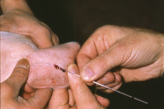
Marginal ear vein
4. Horse: Jugular vein

5. Cats: Jugular vein and medial saphenous vein


Jugular vein


Saphenous vein
6. Dog: Jugular vein and cephalic vein
Jugular vein
Cephalic vein
7. Poultry: Wing vein
Wing vein bleeding
8. Elephant: auricular (ear) vein, cephalic vein, saphenous vein

Saphenous vein

Auricular vein
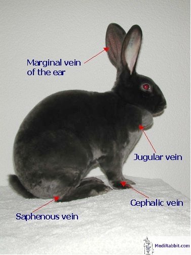
9. Rabbit, rat or mice : Marginal ear vein, jugular vein, cephalic vein, saphenous vein
10. Iguana (or some cold-blooded animal) : Ventral tail vein
Ps: When time comes, I'll post the blood sampling procedure