


My case study
***************************************************

See the lump?

see something here

and again
History
Id: Hay (has been given birth to a calf recently)
Species: Cattle
Age: About 2 and half years
Breed: Charlois
Sex: Female
Weight: About 600 kg
Nutrition: Feed on grass, free grazing
Previous treatment: Deworming
*No vaccination is given

Hay
General inspection
Overall inspection: Healthy
Temperament: Aggressive
Body condition: 5/9
Skin condition: Not smooth, lumps are found throughout the body. Ticks are found engorged on the skin

Ticks isolated from the cow's skin
*Other than that, other findings were consider normal
Physical examination
Temp: 39.1C (quite normal eh?)
Heart rate: 67 bpm
Respiratory rate: (umm..guess i missed this out lol)
Mucous membrane: Pink
TPR: <2second
Skin palpation reveals there were small lump on the skin and many ticks are found within the region. Other findings are normal
Samples taken:
Fecal sample (check on endoparasite)
Tick sample

Me preparing for fecal sample

When taking the fecal sample

A tick
Treatment given: 30 mL Ketasol (vitamin B) IM, Oxytetracyline 1mg/kg IM, Bayticol (cattle tickicide 20mL/cow) pour on method

Me giving Bayticol by pour on method
oh my! i look so fat :(

Bayticol
The next day, I did fecal egg count by using McMaster technique but the result is negative. Maybe it is due to deworming activity done by the farmer. I also checked on the tick and it is positively from the Boophilus species. Since we're not taking any blood sample, so we are not sure weather the cattle is infected with some blood parasite such as Babesiosis or anaplasmosis.
So I did some research to find out about Boophilus tick, of which something I have learnt in parasitology class from the previous semesters. Just for quick revision.

Rhipicephalus (Boophilus) microplus
R. microplus (formely known as Boophilus microplus) is a hard tick that can be found on many hosts including cattle, buffalo, horses, donkeys, goats, sheep, deer, pigs, dogs and some wild animals. Heavy tick burdens on animals can decrease production and damage hides. R. microplus can also transmit babesiosis (caused by the protozoal parasites Babesia bigemina and Babesia bovis) and anaplasmosis (caused by Anaplasma marginale). Under experimental conditions, this tick can transmit Babesia equi, the cause of equine piroplasmosis.
Species affected
R. microplus mainly infests cattle, deer and buffalo, but it can also be found on horses, goats, sheep, donkeys, dogs, pigs and some wild mammals. Cattle are particularly vulnerable when they first encounter cattle ticks but develop a degree of resistance after repeated exposure. Bos indicus cattle (tropical breeds) and their crosses develop better resistance than do Bos taurus (British and European breeds). Horses, goats and sheep also suffer tick-worry but after a period of time they will develop strong resistance.
Life cycle
The cattle tick spends the parasitic stage of its life on the one host (one host tick). This stage takes approximately 21 days during which time the tick changes from a minute larvae to a nymph and finally an adult. Adult females feed slowly for about a week before rapidly filling with blood just prior to detachement. They then drop onto pasture, lay up to 3000 eggs and die. Eggs hatch to produce larvae which infest the pasture until picked up by a suitable host or they die. This non-parasitic stage can vary from approximately two months in summer to six to seven months over winter and is adversely affected by extremes in temperature and moisture levels. Males feed occasionally, but do not fill with blood. They wander over the beast for two months or more, mating with females.

Life cycle R.microplus
Pathology (general)
Ticks are primarily parasites of wild animals and only 10% of species feed on domestic animals, primarily sheep and cattle. The effect of ticks on host species can be divided into:
- Cutaneous effects: Inflammation and infection
- Systemic effects: Transmission of microorganism from another host, paralysis of the hist and bacteraemia resulting of the introduction of micro-organism
At the site of a tick focal dermal necrosis and haemorrhage occur, followed by an inflammatory response often involving eosinophils. Although a hypersensitivity reaction may be involved in the local response, the innate inflammatory response and dermal necrosis is sufficient to damage the hide. Tick bite wounds can become infected with staphylococcus bacteria causing local cutaneous abscesses or pyaemia. Heavy tick infestation can result in significant blood loss, reduced productivity, reduced weight gain and can cause restlessness. Tick bite lesions may also predispose animals to myiasis
Systemis effects: Vectors of disease.
Through their blood feeding habits, ticks are important as vectors of animal disease, transmitting a wide range of pathogenic viruses, rickettsia, bacteria and protozoa. In addition, many of the major diseases transmitted by ticks, such as tick-borne encephalitis, lyme disease, relapsing fever or Rocky Mountain spotted fever are pathogenic to humans. Wild and domestic animals are particularly important as reservoirs of the organisms causing these disease through animal/tick.human cycle of contact.
Ticks are effective vectors because;
- They attach securely to their hosts, allowing them to be transferred to new habitats while on the host
- The lengthy feeding period allows large numbers of pathogens to be ingested
- While feeding the ticks often regurgitate, introducing pathogens to the host
- Many species of tick are long lived
- Females lay large numbers of eggs and therefore tick populations have rapid potential for increase
- Of they fail to fond a host they can survive for lengthy periods without feeding
- Ingested pathogens may be passed from larva to nymph and nymph to adult females to the next generation via ovaries
- Non viraemic transmission may occur between co-feeding ticks
Identification of R.microplus
Rhipicephalus microplus is a member of the family Ixodidae (hard ticks). This tick was formerly known as Boophilus microplus; however, Boophilus has recently become a subgenus of the genus Rhipicephalus. Hard ticks have a dorsal shield (scutum) and their mouthparts (capitulum) protrude forward when they are seen from above. Boophilus ticks have a hexagonal basis capitulum. The spiracular plate is rounded or oval and the palps are very short, compressed, and ridged dorsally and laterally. Males have adanal shields and accessory shields. The anal groove is absent or indistinct in females, and faint in males. There are no festoons or ornamentation.
R. microplus adults have a short, straight capitulum. The legs are pale cream and there is a wide space between
first pair of legs and the snout. The body is oval to rectangular and the shield is oval and wider at the front. The
snout is short and straight. The nymphs of this species have an orange-brown scutum. The body is oval and wider at front. The body color is brown to blue-gray, with white at the front and sides. R. microplus larvae have a short, straight capitulum and a brown to cream body. Larvae have six legs instead of eight.
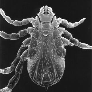
Male
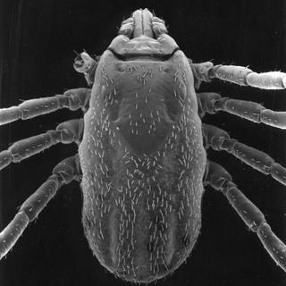
Female

Female (engorged ticks)
Tick control
In regions where this tick is endemic, control methods include acaricide treatment, pasture rotation, environmental modification, and integrated biologic and chemical control strategies. Acaricide resistance is common in the Boophilus subgenus of ticks. The use of resistant breeds is an important means of tick control in some countries. European (Bos taurus) breeds of cattle usually remain fairly susceptible to ixodid ticks, even after multiple exposure.
Ideally, the cattle should be removed from the infected pasture (although this is not always practical) and topical therapy is the used to reduce the tick population. The life cyle of the particular ticks involved will influence the application regime, with multiple-host ticks requiring prolonged insecticidal programmes, whereas control of one-host ticks may only require treatment for a few weeks of the year. Organophosphates and pyrethroids have been used to control tick populations. Resistannce of various topical insecticides is also an increasing problem.
The following compounds have been used in the control of ticks in cattle: Amitraz, chlorpyrifos, cypermectin, cyprothrin, deltamethrin, diazinon, dichlorvos, dioxathion, flumethrin, malathion, permethrin, phosmet, propetamhos and trichlorfon. These are available in various formulations including sprays, dip and slow-release ear tags. Systemic treatment with 200 ug/kg ivermectin every 2-4 weeks has also been shown to prevent tick engorgement and reproduction. Management measures include separation if cattle from infected pasture and cultivation of the infested land.
Sources: Cattle tick, Queensland Government, Rhipicipalus microplus, the center for food security and public health, Vet ectoparasite, biology, pathology, and control 2nd ed Blackwell science.


