Powered by Blogger.
RSS

Welcome

I created this blog as an instrument of what I have encountered in the world of veterinary medicine as a proud vet student. Comments and suggestions are welcome here at;

sweet_daffodil90@yahoo.co.uk

Regards,
Aina Meducci 2012

Disclaimer

The following blog posts is not genuinely from my research but through readings and citation from trusted website. I do not own any of the copyright and therefore you may use it at your own risk

SINCE I AM NOT A VETERINARIAN YET, THEREFORE I CAN'T CONSULT ANY MEDICAL ADVICE TO YOU AND YOUR PETS! EXTREMELY IMPORTANT!.

Happy reading!
Post Icon

Hyperkalemia

While still struggling to understand on fluid therapy, my attention were hooked on hyperkalemia.

************************************************************


Hyperkalemia is an excessive level of potassium in the bloodstream. Potassium has several important functions in the body. It is essential for the normal functioning of the muscles, heart, and nerves. Potassium helps the body regulate activity of muscle, including the smooth muscle (involuntary muscles, such as the muscles found in the digestive tract), skeletal muscle (voluntary muscles, such as muscles of the extremities and torso), and the muscle of the heart. It is also important for maintaining normal heart electrical rhythm and for normal electrical signals in the nervous system.


Extreme hyperkalemia is a medical emergency due to the risk of potentially fatal abnormal heart rhythms (arrhythmia).


  • The normal potassium level in the blood is 3.5-5.0 milliEquivalents per liter (mEq/L).

  • Potassium levels between 5.1 mEq/L to 6.0 mEq/L are considered to be mild hyperkalemia.

  • Potassium levels of 6.1 mEq/L to 7.0 mEq/L are moderate hyperkalemia, and levels above 7 mEq/L reflect severe hyperkalemia


Normal Potassium Physiology

Potassium is critical for the normal functioning of the muscles, heart, and nerves. It plays an important role in controlling activity of smooth muscle (such as the muscle found in the digestive tract) and skeletal muscle (muscles of the extremities and torso), as well as the muscles of the heart. It is also important for normal transmission of electrical signals throughout the nervous system within the body.

Normal blood levels of potassium are critical for maintaining normal heart electrical rhythm. Both low blood potassium levels (hypokalemia) and high blood potassium levels (hyperkalemia) can lead to abnormal heart rhythms.

The most important clinical effect of hyperkalemia is related to electrical rhythm of the heart. While mild hyperkalemia probably has a limited effect on the heart, moderate hyperkalemia can produce EKG changes (EKG is an electrical reading of the heart muscles), and severe hyperkalemia can cause suppression of electrical activity of the heart and can cause the heart to stop beating.

Another important effect of hyperkalemia is interference with functioning of the skeletal muscles. Hyperkalemic periodic paralysis is a rare inherited disorder in which patients can develop sudden onset of hyperkalemia which in turn causes muscle paralysis. The reason for the muscle paralysis is not clearly understood, but it is probably due to hyperkalemia suppressing the electrical activity of the muscle.


Normal rhythm of the heart


Hyperkalemia rhythm of heart


Causes of hyperkalemia

The major causes of hyperkalemia are kidney dysfunction, diseases of the adrenal gland, potassium sifting out of cells into the blood circulation, and medications.

The majority of potassium within the body (about 98%) is located within cells, with only 2% located in the bloodstream. A number of conditions can cause potassium to move out of the cells into the blood circulation, thereby increasing the measured level of potassium in the blood, even though the total amount of potassium in the body has not changed.

Similarly, any condition in which there is massive tissue destruction can result in elevated levels of blood potassium as the damaged cells release their potassium. Examples of tissue destruction include:

  • Trauma
  • burns,
  • Surgical procedures,
  • Destruction of tumor cells or red blood cells
  • Rhabdomyolysis (a condition involving destruction of muscle cells that is sometimes associated with muscle injury, alcoholism, or drug abuse).

Moreover, difficulty in drawing blood from veins for testing can traumatize red blood cells, releasing potassium into the serum of the blood sample to cause a falsely elevated reading of hyperkalemia on the blood test.

Any condition that decreases kidney function can result in hyperkalemia, since the kidneys rid the body of excess potassium by excreting it in the urine. Examples of conditions that decrease kidney function are

  • glomerulonephritis,
  • acute or chronic renal failure,
  • transplant rejection
  • obstructions within the urinary tract (such as the presence of stones)

The adrenal glands secrete many hormones important for proper body function. Among these is aldosterone, which regulates the retention of sodium and fluid in the kidneys along with the excretion of potassium in the urine. Diseases of the adrenal gland (such as Addison's disease, that causes a decreased aldosterone secretion) lead to a decrease in kidney excretion of potassium resulting in hyperkalemia.

Examples of medications that may lead to elevated potassium levels include:

  • Nonsteroidal antiinflammatory drugs (NSAID)
  • ACE inhibitors,
  • Angiotensin II receptor blockers (ARBs),
  • Diuretics.

Hyperkalemia Symptoms

  • Irregular heartbeat
  • Nausea
  • Slow, weak pulse

Diagnosis

ECG test

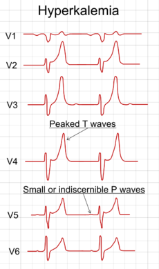
With mild to moderate hyperkalemia, there is reduction of the size of the P wave and development of peaked T waves. Severe hyperkalemia results in a widening of the QRS complex, and the EKG complex can evolve to a sinusoidal shape. There appears to be a direct effect of elevated potassium on some of the potassium channels that increases their activity and speeds membrane repolarization.

Also, (as noted above), hyperkalemia causes an overall membrane depolarization that inactivates many sodium channels. The faster repolarization of the cardiac action potential causes the tenting of the T waves, and the inactivation of sodium channels causes a sluggish conduction of the electrical wave around the heart, which leads to smaller P waves and widening of the QRS complex.

Abnormal rhythm of the heart


Treatment of hyperkalemia

When arrhythmias occur, or when potassium levels exceed 6.5 mmol/l, emergency lowering of potassium levels is mandated. Several agents are used to transiently lower K+ levels. Choice depends on the degree and cause of the hyperkalemia, and other aspects of the patient's condition.


Treatment for acute hyperkalemia

1) Calcium chloride or calcium gluconate

Calcium (Calcium chloride or calcium gluconate) increases threshold potential through a mechanism that is still unclear, thus restoring normal gradient between threshold potential and resting membrane potential, which is elevated abnormally in hyperkalemia. One ampule of Calcium chloride has approximately 3 times more calcium than calcium gluconate. Onset of action is <5 min and lasts about 30-60 min. Doses should be titrated with constant monitoring of ECG changes during administration and the dose should be repeated if ECG changes do not normalize within 3 to 5 min.

2) Insulin

Insulin (e.g. intravenous injection of 10-15 units of regular insulin along with 50ml of 50% dextrose to prevent hypoglycemia) will lead to a shift of potassium ions into cells, secondary to increased activity of the sodium-potassium ATPase. Its effects last a few hours, so it sometimes needs to be repeated while other measures are taken to suppress potassium levels more permanently.


3) Albuterol

Albuterol, Ventolin) is a β2-selective catecholamine that is administered by nebulizer (e.g. 10–20 mg). This drug also lowers blood levels of K+ by promoting its movement into cells.


4) Furosemide (diuretics)

Increase elimination of potassium to the urine

5) Polystyrene sulfonate with sorbitol (Kayexalate) either orally or rectally is widely used with the goal to lower potassium over several hours. Removal of potassium is assumed to require defecation. However, careful clinical trials to demonstrate the effectiveness of Kayexalate are lacking, and there are small risks of necrosis of the colon.


Long term prevention

Preventing recurrence of hyperkalemia typically involves reduction of dietary potassium,removal of an offending medication, and/or the addition of oral bicarbonate or a diuretic (such as furosemide or hydrochlorothiazide). Polystyrene sulfonate and sorbital (Kayexalate) is occasionally used on an ongoing basis to maintain lower serum levels of potassium.

Sources: Wikipedia.net, Hyperkalemia, AAPF, Hyperkalemia, Medline Plus, hyperkalemia, emedicine.net,


  • Digg
  • Del.icio.us
  • StumbleUpon
  • Reddit
  • RSS
Post Icon

Fluid therapy

What a stressful week.


********************************************************


Fluid therapy in dogs


Fluid Therapy is the administration of fluids to a patient as a treatment or preventative measure. It can be administered via an intravenous, intraperitoneal, intraosseous, subcutaneous and oral routes. 60% of total bodyweight is accounted for by the total body water. This can further be divided into intracellular or extracellular as shown below.


Body Compartments.jpg


Fluid therapy is indicated either when there is a loss of fluid to any part of these compartments or there is a risk of loss of fluid. The severity of the fluid loss, and the compartment from which it has been lost, influence the choice of fluid and the speed at which it needs to be administered. If fluid therapy is performed as a treatment then it is necessary to diagnose and treat the underlying condition.


Indications

  • Hypotension
  • Hypovolemia
  • Electrolyte, metabolic and acid base disorders
  • Decreased oxygen delivery
  • Geriatric patients at risk of organ failure

Degree of dehydration in animal

5% (below): No physical finding
5%: Dry oral mucous membrane but no panting or pathological bradycardia
7% : Mild moderate decreased skin turgor, dry oral mucous membranes, slight tachycardia,
10%:Moderate to marked degree of decreased skin turgor, dry oral mucous membrane, tachycardia, decrease pulse pressure
12%: Marked loss of skin turgor, dry oral mucous membrane and significant of shock


In mild dehydration, subcutaneous fluids are useful. Isotonic fluids should be used and no more than 5 to 10 ml/lb should be given at each injection site. The rate of subcutaneous fluid flow usually is governed by patient comfort. These fluids are aseptically administered and multiple sites are required to provide adequate fluid volume. Generally, all subcutaneous fluids are resorbed within 6 to 8 hours. If fluids are still noted subcutaneously after this time, the use of intravenous fluids to reestablish peripheral perfusion should be considered.


subcutaneous fluid cat


The intraperitoneal route is quick, easy, and the fluids will generally be reabsorbed thus increasing the circulating volume. However, there is the potential of bacterial peritonitis, perforating viscera, and decreasing ventilation from impeding diaphragmatic excursion. Experience with peritoneal dialysis in dogs has shown that peritoneal fluids often traverse the diaphragm, entering the thoracic space, and further affecting ventilation. Currently, intraperitoneal fluids cannot be recommended.


In general, intravenous fluid administration is indicated in dogs and cats with 7% or greater dehydration. There are numerous potential routes for intravenous fluid administration:

  • Peripheral veins
  • Jugular veins
  • Intraosseous

The amount of fluid needed for replacement depends on the patient's status. Of primary concern is the status of the blood volume and later concern is directed to restoration of total body water and electrolytes

3 phases of fluid therapy

  • Emergency phase
  • Replacement phase
  • Maintenance phase

Types of fluid

1. Crystalloid


Crystalloid fluid

A crystalloid fluids form a true solution meaning it can easily cross a semi permeable membrane and is distinguished by its ability to be crystallised. They can be classified asmaintainance or replacement fluids. A replacement crystalloid often do not have high potassium concentrations allowing for rapid administration without potassium toxicity, where as maintainance crystalloids often have a higher potassium but lower sodium and chloride then a replacement crystalloid.

0.9% Sodium Chloride

Also known as normal saline, 0.9% sodium chloride is often used as a replacement solution. It contains no further electrolytes. It has a higher chloride concentration then plasma. This can lead to a hyperchloremic acidosis which is unlikely to cause any problems in healthy patients but may cause further issues in compromised patients.


Hartmanns/Lactated Ringers

Hartmanns or Lactated Ringers Solution (LRS) is a balanced electrolyte solution. It also contains lactate which aids correction of acidosis. It also contains potassium and calcium. It also has a lower sodium concentration then plasma. Due to the calcium, it is not possible to administer LRS with blood products or sodium bicarbonate as it leads to clot/crystal formation. It is commonly used as a maintenance solution.


5% Dextrose

5% Dextrose consists of dextrose in water, and no electrolytes. It is rarely indicated for use during surgeries. It should not be used in patients thought to have cerebral injuries due to the deterimental affects the glucose will have.


2. Colloid solution

A colloid solution contains large molecules that do not easily cross a semi permeable membrane. Colloids can be either natural or synthetic. They are used to help maintain colloid osmotic pressure (COP), correct hypovolemia and with plasma (see below) clotting factors. It is the number of particles within the colloid that influences the osmotic effect, not the size of the particles. They are highly efficient at expanding vascular volume, compared to crystalloids, which easily diffuse out of the circulation, meaning that less volume is required

Dextran

Dextran is a molecule produced by certain strains of bacteria to give this linear polysaccaharide as either a high or low weight molecule. It is found in a sodium chloride or dextrose solution. It is metabolised to glucose within the patient. Both high and low weight molecules produce the same plasma volume expansion per gram, although the low weight molecule it thought to achieve this more rapidly. There are concerns about it's interaction with fibrin, making clots weaker and decreasing factor VIII and von Willebrand's factor.Examples include Dextran 40 and 70.


Hetastarch

Hetastarch is a synthetic glucose polymer, most commonly in a sodium chloride solution. Again, it is broken down into glucose in the patient. It is not as long in duration of action as dextran products but is still a very effective plasma volume expander. It can be used to draw fluid out of intersitium in patients with peripheral oedema or ascites. Again, there is some concern about prolonged bleeding times in patients who have received hetastarch products.Examples include Voluven.


Gelatin Solution

Gelatin solutions are derived from degradation of bovine collagen. It is found in several forms. Their advantage over other colloid solutions are their minimal effects on coagulation and minimal antigenic reactions. They have a lower molecular weight also.Examples include Haemaccel.


Blood Products

Whole Blood


Whole blood is administered when a patient requires all the components of whole blood. It contains clotting factors and active platelets and so requires correct storage. It is important to monitor patients receiving blood products closely in case of an anaphylactic reaction. Another concern with whole blood is the effect of citrate on the availability of calcium, which may require dosing also through a different line.

Plasma

Plasma is available as either a fresh or frozen product. Fresh plasma contains platelets and clotting factors, while fresh frozen plasma contains no platelets but does have clotting factors as long as it has been thawed correctly. Fresh frozen plasma can be used to expand the plasma volume, as long as the packed cell volume of that patient is within normal limits. However, there is always a concern when administering blood products of an anaphylactic reaction and so patients receiving these products should be closely monitored.


Fluid rate calculation

When calculating the fluid requirements of a patient, there are 3 elements to consider

  • Replacement
  • Maintainance
  • Ongoing Losses

For example of calculation refer here


Monitoring fluid therapy

Fluid therapy can be monitored by observing urine output, Packed Cell Volume (PVC), Total Protein (TP, or Total Solids, TS), mucous membrane colour, and blood pressure among others. It is important to also observe for signs of oedema, such as pulmonary oedema or ascites. It should not be stopped until hydration of the patient has returned to normal and the patient is able to maintain a normal hydration status independently.


Special consideration

  • Shock-require immediate fluid therapy (crystalloid)
  • Anesthesia (pre, intra, post) -Warmed fluid to combat hypothermia
  • Cardiac disease -avoid fluid overload
  • Hepatic disease
  • CNS disease-Avoid giving glucose-contained fluid

Sources: Fluid therapy;wikivet, fluid and electrolyte imbalance www.cvmbs.colostate.edu

  • Digg
  • Del.icio.us
  • StumbleUpon
  • Reddit
  • RSS
Post Icon

Anesthesiology: Stages of anesthesia


I have many things to be posted here, but the time seems to be unkind, I need to steal time in order for me to write freely here, without interruptions.

** I've got to beat the time!

***********************************************************

General anesthesia


General anesthesia (GA) causes total loss of sensation, and complete loss of consciousness in the patient. They are administered by inhalation of certain gases or vapourized liquids, intravenous infusion, or rectal induction.

Example of inhalant anesthetics: Isoflurane, Nitrous oxide, desflurane, halothane, thiopental


The induction of inhalant anesthesia is divided into 4 stages.


Stage 1

Refers as stage of analgesia (reduce pain sensation) and induction (conscious to unconciousness). During this period, patient experiences dizziness, sense of unreality, and less sensitivity of pain. Patients sense of hearing is increased and and response to noises are intensified.


Stage 2

Stage of excitement. During this period, there is a variety of reactions involving muscular activity and delirium. Patient may become very aggressive. All the reflexes still present and may appear exaggerated. Patient may exhibit involuntary excitement in the firm of rapid movement of the limbs, vocalisation and struggling.


Stage 3

Surgical and operative stage. It is subdivided into 4 planes (Plane 1- plane 4)

Plane i

  • Respiratoty pattern becomes regular
  • Involuntary movement cease
  • Eyeball rotates ventrally
  • Pupils may be partially constricted
  • Depressed swallowing reflexes (to insert endotracheal tube)

Plane ii

  • Increased heart rate and respiration rate
  • Patients unconscious and immobile
  • Eyeball central or ventral
  • Skeletal muscle become relax
  • Protective reflexes lost

Plane iii

  • Patient deeply anesthetised
  • Pulse strength reduce due to reduce blood pressure
  • Increase capillary refill time
  • Eyeball become central
  • Pupils moderately dilated
  • Absent reflex activity
  • Skeletal muscle relaxation
Plane iv

  • Rocking ventilation
  • Decrease ventilation
  • Fully dilated pupils
  • Absence of pupillary reflex
  • Eyes become dry due to absence of lacrimal secretion
  • Muscle tome flaccid
  • Depression of CVS (drop heart rate and blood pressure)
  • Pale mucous membrane
  • Prolonged CRT
This stage is in danger in respiratory and cardiac arrest.


Stage 4

**Due to increase past stage 3!!

  • Cessation or respiration (skeletal muscle too relax)
  • Circulatory collapse
  • DEATH!
**Immediate resuscitation is necessary to save patient's life


Recovery (not include in the stage of anesthesia)

Begins when the concentration anesthetics agent in brain decrease (varies in anesthetic agents).


Summary of stages of anesthesia


Sources; Veterinary anesthesia and analgesia 3rd edition, Mosby publication, General anethesia www.tpub.com


  • Digg
  • Del.icio.us
  • StumbleUpon
  • Reddit
  • RSS
Post Icon

Diabetes


In surgery class, we often talk about causes of gangrene which are common to diabetic people. So here's the interesting part: untreated or superinfection wounds maybe lead to lower extremities amputation!


*******************************************************************


A diabetic cat suffer from feline diabetes
(is this pic true?)


Diabetes mellitus, often simply referred to as diabetes, is a group of metabolic diseases in which a person has high blood sugar (hyperglycemia), either because the body does not produce enough insulin, or because cells do not respond to the insulin that is produced. This high blood sugar produces the classical symptoms of polyuria (frequent urination), polydipsia (increased thirst) and polyphagia (increased hunger).

Diabetes insipidus is a condition in which the kidneys are unable to conserve water. It is characterized by excessive thirst and excretion of large amounts of severely diluted urine, with reduction of fluid intake having no effect on the concentration of the urine. There are several different types of DI, each with a different cause. The most common type in humans is central DI, caused by a deficiency of arginine vasopressin (AVP), also known as antidiuretic hormone(ADH). The second common type of DI is nephrogenic diabetes insipidus, which is caused by an insensitivity of the kidneys to ADH. It can also be an iatrogenic artifact of drug use.


Although they have a common name, diabetes mellitus and diabetes insipidus are two entirely separate conditions with unrelated mechanisms. Both cause large amounts of urine to be produced (polyuria), and the term diabetes is derived from the Greek name for this symptom. However, diabetes insipidus is either a problem with the production of antidiuretic hormone (cranial diabetes insipidus) or kidney's response to antidiuretic hormone (nephrogenic diabetes insipidus), whereas diabetes mellitus causes polyuria via a process called osmotic diuresis, due to the high blood sugar leaking into the urine and taking excess water along with it.


I will only discuss on Diabetes mellitus**


There are three main types of diabetes:


Type 1 diabetes

Results from the body's failure to produce insulin, and presently requires the person/animal to inject insulin. (Also referred to as insulin-dependent diabetes mellitus, IDDM for short, and juvenile diabetes.) Type 1 diabetes mellitus is characterized by loss of the insulin-producing beta cells of the islets of Langerhans in the pancreas leading to insulin deficiency. This type of diabetes can be further classified as immune-mediated or idiopathic. The majority of type 1 diabetes is of the immune-mediated nature, where beta cell loss is a T-cell mediated autoimmuneattack.It can occur at any age, but it is most often diagnosed in children, teens, or young adults. In this disease, the body makes little or no insulin. Daily injections of insulin are needed.

Type 2 diabetes

Results from insulin resistance, a condition in which cells fail to use insulin properly, sometimes combined with an absolute insulin deficiency. (Formerly referred to as non-insulin-dependent diabetes mellitus, NIDDM for short, and adult-onset diabetes.)

The pancreas secretes insulin, but the body is partially or completely unable to use the insulin. This is sometimes referred to as insulin resistance. The body tries to overcome this resistance by secreting more and more insulin. People with insulin resistance develop type 2 diabetes when they do not continue to secrete enough insulin to cope with the higher demands.

Type 2 diabetes is typically recognized in adulthood, usually after age 45 years. It used to be called adult-onset diabetes mellitus, or non-insulin-dependent diabetes mellitus. These names are no longer used because type 2 diabetes does occur in younger people, and some people with type 2 diabetes need to use insulin.It is usually controlled with diet, weight loss, exercise, and oral medications. Some of the causes include; high blood pressure, high blood fat level and overweight.

Gestational diabetes

Is when pregnant women, who have never had diabetes before, have a high blood glucose level during pregnancy. It may precede development of type 2 DM.


Pathophysiology of Diabetes Mellitus


Sign and Symptoms

In susceptible animal such as cats and dogs, polyurination are common cause. The cat may also drink excessive of water. Feline patients are urinating vast amounts, because the water gets literally pulled out of the organism through the osmotic effect of the large sugar molecules. So they need to replace this water by drinking a lot.

Weight loss is another specific sign. Fact is, that a diabetic cat eats more than a healthy animal. The high blood sugar level in the blood stream initiates the brain to signal the body to eat even more to be able to get the blood sugar into the body tissues.If this cat illness is present for a longer time, a cat becomes eventually lethargic and very sick.

Diabetic Cat Walking on Hock

A fairly common problem in diabetic cats is a hindlimb weakness that may be caused by the increased blood sugar level.Constantly high glucose in the bloodstream causes conductivity problems in peripheral nerve axons, especially in the tibial nerve of one or both sides.

Typical signs are walking on the hocks or a 'plantigrade stance' and a weakness in the hindlimbs of variable severity.

And these are human symptoms;


Diabetes complications

Both forms of diabetes ultimately lead to high blood sugar levels, a condition called hyperglycemia. Over a long period of time, hyperglycemia damages the retina of the eye, the kidneys, the nerves, and the blood vessels.Damage to the retina from diabetes (diabetic retinopathy) is a leading cause of blindness.

  • Damage to the kidneys from diabetes (diabetic nephropathy) is a leading cause of kidney failure.

  • Damage to the nerves from diabetes (diabetic neuropathy) is a leading cause of foot wounds and ulcers, which frequently lead to foot and leg amputations.

  • Damage to the nerves in the autonomic nervous system can lead to paralysis of the stomach (gastroparesis), chronic diarrhea, and an inability to control heart rate and blood pressure during postural changes.

  • Diabetes accelerates atherosclerosis, (the formation of fatty plaques inside the arteries), which can lead to blockages or a clot (thrombus). Such changes can then lead to heart attack, stroke, and decreased circulation in the arms and legs (peripheral vascular disease).

  • Diabetes predisposes people to high blood pressure and high cholesterol and triglyceride levels. These conditions independently and together with hyperglycemia increase the risk of heart disease, kidney disease, and other blood vessel complications.

In the short run, diabetes can contribute to a number of acute (short-lived) medical problems.

Many infections are associated with diabetes, and infections are frequently more dangerous in someone with diabetes because the body's normal ability to fight infections is impaired. To compound the problem, infections may worsen glucose control, which further delays recovery from infection.

Hypoglycemia, or low blood sugar, occurs from time to time in most people with diabetes. It results from taking too much diabetes medication or insulin (sometimes called an insulin reaction), missing a meal, doing more exercise than usual, drinking too much alcohol, or taking certain medications for other conditions. It is very important to recognize hypoglycemia and be prepared to treat it at all times. Headache, feeling dizzy, poor concentration, tremors of hands, and sweating are common symptoms of hypoglycemia. You can faint or have a seizure if blood sugar level gets too low.

Diabetic ketoacidosis is a serious condition in which uncontrolled hyperglycemia (usually due to complete lack of insulin or a relative deficiency of insulin) over time creates a buildup in the blood of acidic waste products called ketones. High levels of ketones can be very harmful. This typically happens to people with type 1 diabetes who do not have good blood glucose control. Diabetic ketoacidosis can be precipitated by infection, stress, trauma, missing medications like insulin, or medical emergencies like stroke and heart attack.

Hyperosmolar hyperglycemic nonketotic syndrome is a serious condition in which the blood sugar level gets very high. The body tries to get rid of the excess blood sugar by eliminating it in the urine. This increases the amount of urine significantly and often leads to dehydration so severe that it can cause seizures, coma, and even death. This syndrome typically occurs in people with type 2 diabetes who are not controlling their blood sugar levels, who have become dehydrated, or who have stress, injury, stroke, or are taking certain medications, like steroids.


Diabetic foot ulcer (my favorite part!!)

Diabetic foot ulcers (an open wound or sore that develops on the skin) are one of the major causes of gangrene and amputation in people/animal with diabetes. Gangrene is results by
  • reduced sensation
  • reduced blood supply
These are described in more detail below.

Reduced sensation

The high blood sugar that is associated with diabetes can cause damage to the nerves (peripheral neuropathy), particularly in feet. Once damaged, the nerves are unable to transmit sensations of pain to your brain.Therefore it is easy to damage or injure your foot by treading on something sharp, or to develop a blister due to ill-fitting shoes without actually realising it.

As a result of the lack of pain, animal may continue walking without protecting the wound. This can make the wound worse and it may develop into an ulcer.

Reduced blood supply

High blood sugar can damage your blood vessels, causing the blood supply to feet to become restricted. If the skin on feet receives less blood, it will also receives a lower number of infection-fighting cells, which means that wounds will take longer to heal.Therefore, the reduced sensation means that animals are more likely to develop an ulcer, and the reduced blood supply means that the ulcer is more likely to become infected. The infection is likely to restrict the blood supply further, leading to gangrene. Worse gangrene may lead to amputation to prevent spreading of bacteria from the rest of the body.

**If person/animal have type 1, or type 2, diabetes, it is essential that to take extra care of feet.


Gangrene


Prevention and treatment

Self-treatment
  • Keep an ideal weight
  • Healthy lifestyle
  • Exercise
So here's the cycle

Medical treatment

Type 1 DM

  • Insulin via injection. Orally taken may destroy the insulin in the stomach
  • Eating healthy diet

Type 2 DM

  • Sulfonylurea or biguanide [metformin Glucophage)], to help control blood sugar level.
  • Insulin injection-control sugar level
  • It is becoming more common for people with type 2 diabetes to take a combination of oral medication and insulin injections to control blood sugar levels.

Medications

Sulfonylureas: These drugs stimulate the pancreas to make more insulin.

Biguanides: These agents decrease the amount of glucose produced by the liver.

Alpha-glucosidase inhibitors: These agents slow absorption of the starches one eats. This slows down glucose production.

Thiazolidinediones: These agents increase sensitivity to insulin.

Meglitinides: These agents stimulate the pancreas to make more insulin.

D-phenylalanine derivatives: These agents stimulate the pancreas to produce more insulin more quickly.

Amylin synthetic derivatives: Amylin is a naturally occurring hormone secreted by the pancreas along with insulin. An amylin derivative, such as pramlintide (Symlin), is indicated when blood sugar control is not achieved despite optimal insulin therapy. Pramlintide is administered as a subcutaneous injection along with insulin and helps achieve lower blood sugar levels after meals, helps reduce fluctuation of blood sugar levels throughout the day, and improves hemoglobin A1C levels.

Incretin mimetics: Incretin mimetics promote insulin secretion by the pancreas and mimic other blood sugar level lowering actions that naturally occur in the body.


Sources: Wikipedia, Gangrene www.nhs.uk, Diabetic in cats www.pet-health-pro.com, Diabetes, pubmed health, Diabetes emedicinehealth.com


  • Digg
  • Del.icio.us
  • StumbleUpon
  • Reddit
  • RSS
Post Icon

Edema

What a day! Pathology seems to be the difficult subject to study, and it is a subject where you need to be extraordinary good in histology and physiology. Plus, when you forgot something what you learned during biology class, u be better say bye2 to pathology!

********************************************************


Ascites (swelling of peritoneal area)


Edema refers to the swelling of a tissue that results from excessive accumulation of fluid within the tissue. Edema can be highly localized as occurs in a small region of the skin subjected to a bee sting. Edema, however, can also comprise an entire limb, specific organs such as the lungs (e.g., pulmonary edema) or the whole body.


General principle of edema

There are two primary fluid compartments in the body between which fluid is exchanged - the intravascular and extravascular compartments.The intravascular compartment contains fluid (i.e., blood) within the cardiac chambers and vascular system of the body. The extravascular system is everything outside of the intravascular compartment. Fluid and electrolytes readily move between these two compartments. The extravascular compartment is made up of many subcompartments such as the cellular, interstitial, and lymphatic subcompartments, and a specialized system containing cerebrospinal fluid in the central nervous system.


The movement of fluid and accompanying solutes between compartments (mostly water, electrolytes, and small molecular weight solutes) is governed by physical factors such as hydrostatic and oncotic forces. These forces are normally balanced in such a manner that fluid volume remains relatively constant between the compartments. If the physical forces or barriers to fluid movement are altered, the volume of fluid may increase in one compartment and decrease in another. In some cases, total fluid volume increases in the body so that both intravascular and extravascular compartments increase in volume. This can occur, for example, when the kidneys fail to excrete sufficient amounts of sodium and water.


When the fluid volume within the interstitial compartment (space between the cells and blood vessels) increases, this compartment will increase in size leading to tissue swelling (i.e., edema). This is what happens when an ankle is sprained and swells. When excess fluid accumulates within the peritoneal space (space between the abdominal wall and organs), this is termed "ascites." Pulmonary congestion, which can occur in heart failure as the left atrial pressure increases and blood backs up in the pulmonary circuit, causes pulmonary edema.


Filtration is the movement of fluid out of the capillary and reabsorption is the movement of fluid back into the capillary. In most capillary systems of the body, there is a small net filtration(typically about 1% of plasma) of fluid from the intravascular to the extravascular compartment. In other words, capillary fluid filtration exceeds reabsorption. This would cause fluid to accumulate within the interstitium (i.e., cause edema) over time if it were not for the lymphatic system that removes excess fluid from the interstitium and returns it back to the intravascular compartment. Therefore, Fluid balance occurs when:

Filtration = Reabsorption + Lymphatic Flow

Circumstances, however, can arise in which net capillary filtration exceeds the capacity of the lymphatics to carry away the fluid (i.e., net filtration > lymph flow). When this occurs, the interstitium will swell with fluid, and thereby become edematous.


capillary fluid filtration and reabsorption


Factors contributed to edema:

  • Increased hydrostatic pressure
  • Reduced oncotic pressure (pressure of plasma protein) in blood
  • Increased tissue oncotic pressure
  • Increased blood vessel permeability (eg Inflammation)
  • Obstruction of lymphatic system
  • Retention of water and sodium in kidney

Classification of edema

1. Subcut edema

Cutaneous edema is referred to as "pitting" when, after pressure is applied to a small area, the indentation persists for some time after the release of the pressure. Peripheral pitting edema, as shown in the illustration, is the more common type, results from water retention. It can be caused by systemic diseases, pregnancy in some women, either directly or as a result of heart failure, or local conditions such as varicose veins, thrombophlebitis, insect bites, anddermatitis.

Non-pitting edema is observed when the indentation does not persist. It is associated with such conditions as lymphedema, Lipoedema and myxedema.


2. Generalize edema

Causes of edema which are generalized to the whole body can cause edema in multiple organs and peripherally. For example, severe heart failure can cause pulmonary edema, pleural effusions, ascites and peripheral edema.


3. Organ specific

Edema will occur in specific organs as part of inflammations, tendonitis or pancreatitis, for instance. Certain organs develop edema through tissue specific mechanisms.

Examples of edema in specific organs:

  • Cerebral edema is extracellular fluid accumulation in the brain. It can occur in toxic or abnormal metabolic states and conditions such as systemic lupus or reduced oxygen at high altitudes. It causes drowsiness or loss of consciousness.

  • Pulmonary edema occurs when the pressure in blood vessels in the lung is raised because of obstruction to remove blood via the pulmonary veins. This is usually due to failure of the left ventricle of the heart. It can also occur in altitude sickness or on inhalation of toxic chemicals. Pulmonary edema produces shortness of breath. Pleural effusions may occur when fluid also accumulates in the pleural cavity.

  • Edema may also be found in the cornea of the eye with glaucoma, severe conjunctivitis or keratitis or after surgery. It may produce coloured haloes around bright lights.

  • Edema surrounding the eyes is called periorbital edema or eye puffiness. The periorbital tissues are most noticeably swollen immediately after waking, perhaps due to the gravitational redistribution of fluid in the horizontal position.

  • Common appearances of cutaneous edema are observed with mosquito bites, spider bites, bee stings (wheal and flare), and skin contact with certain plants such as Poison Ivy orWestern Poison Oak,the latter of which are termed contact dermatitis.

  • Another cutaneous form of edema is myxedema, which is caused by increased deposition of connective tissue. In myxedema (and a variety of other rarer conditions) edema is due to an increased tendency of the tissue to hold water within its extracellular space. In myxedema this is because of an increase in hydrophilic carbohydrate-rich molecules (perhaps mostly hyaluronan) deposited in the tissue matrix. Edema forms more easily in dependent areas in the elderly (sitting in chairs at home or on aeroplanes) and this is not well understood. Estrogens alter body weight in part through changes in tissue water content. There may be a variety of poorly understood situations in which transfer of water from tissue matrix to lymphatics is impaired because of changes in the hydrophilicity of the tissue or failure of the 'wicking' function of terminal lymphatic capillaries.

  • In lymphedema abnormal removal of interstitial fluid is caused by failure of the lymphatic system. This may be due to obstruction from, for example, pressure from a cancer or enlarged lymph nodes, destruction of lymph vessels by radiotherapy, or infiltration of the lymphatics by infection (such as elephantiasis). It is most commonly due to a failure of the pumping action of muscles due to immobility, most strikingly in conditions such as multiple sclerosis, or paraplegia. Lymphatic return of fluid is also dependent on a pumping action of structures known as lymph hearts. It has been suggested that the edema that occurs in some people following use of aspirin-like cyclo-oxygenase inhibitors such as ibuprofen or indomethacin may be due to inhibition of lymph heart action.

  • Hydrops fetalis is a condition of the fetus characterized by an accumulation of fluid, or edema, in at least two fetal compartments.

Treatment of edema

Reduce salt (sodium) in diet — Sodium, which is found in table salt, can worsen edema. Reducing the amount of salt consumed can help to reduce edema.

Diuretics — Diuretics are a type of medication that causes the kidneys to excrete more water and sodium, which can reduce edema. Diuretics must be used with care because removing to much fluid too quickly can lower the blood pressure and impair kidney function.

In heart failure patients, improving cardiac output by using cardiostimulatory or vasodilators drugs reduces venous and capillary pressures, thereby decreasing filtration and promoting reabsorption of fluid within tissues.


Sources: Edema: Wikipedia, CV Physiology: tissue edema, edema (swellling) www.uptodate.com

  • Digg
  • Del.icio.us
  • StumbleUpon
  • Reddit
  • RSS