Powered by Blogger.
RSS

Welcome

I created this blog as an instrument of what I have encountered in the world of veterinary medicine as a proud vet student. Comments and suggestions are welcome here at;

sweet_daffodil90@yahoo.co.uk

Regards,
Aina Meducci 2012

Disclaimer

The following blog posts is not genuinely from my research but through readings and citation from trusted website. I do not own any of the copyright and therefore you may use it at your own risk

SINCE I AM NOT A VETERINARIAN YET, THEREFORE I CAN'T CONSULT ANY MEDICAL ADVICE TO YOU AND YOUR PETS! EXTREMELY IMPORTANT!.

Happy reading!
Post Icon

Final Exam Sem 5 January 2012

Dear fellows,
I may not be available until next January due to final semester 5 exam.
Most of my time will be spent on tests, books and practicals
Wish me good luck!


Final Exam Semester 5

1-14 January 2012


  • Digg
  • Del.icio.us
  • StumbleUpon
  • Reddit
  • RSS
Post Icon

Haemonchosis (Barber's pole worm) infestation

The haemonchosis infestation came out in pathology test today! It reminds me of Veterinary Institute of Malaysia, a year ago doing my voluntary practice in ruminant. We came across this case.

**************************************************


Haemonchos contortus


Haemonchus contortus, also known as red stomach worm, wire worm or Barber's pole worm, is very common parasite and one the most pathogenic nematode of ruminants. H. contortus is a highly pathogenic parasitic nematode of that can infect a large number of wild and domesticated ruminant species and is the most economically important parasite of sheep and goats worldwide. Although originally a tropical parasite, it has been disseminated around the world by livestock movement and can now be found as far north as the arctic circle.

Adult worms are blood feeders that reside in the abomasum (stomach) and are approximately 2cm in length when mature. They are dioecious with single females typically producing several thousand eggs per day which pass out of the host in faeces and develop to infective larvae on the pasture.


H.contortus on the abomasal surface of the infected sheep


Anemia, low packed cell volume (PCV), diarrhea, dehydration,peripheral, and internal fluid accumulation are common signs of barber pole worm infestation. Infested goats have lower growth rates, markedly reduced reproductive performance, and have higher rates of illness and death. Consequently, H.contortus may account for greatly reduced profits in a goat operation.


Fluid accumulation at the submandibular region (bottle jaw)


Life cycle of H.contortus

Goats are born without H. contortus; however,they become infested with the worm when theystart grazing. The L3, or infecting larvae, are eatenby the goats during grazing. The L3 larvae thenburrow into the internal layer of the goat's abomasums (true stomach) where they develop to a L4, or pre–adult larvae. The L4 molts into L5, the adult form.

Adult male and female worms live in the abomasum of goats, where they feed on blood. The worms mate and produce eggs. Adult females deposit from 5,000 to 10,000 eggs per day, whichare passed through goat feces to the pasture. Eggsare hatched either in soil or water. When the soil iswarm and moist, eggs will hatch into L1 larvae(first stage juveniles). The L1 larvae then develop through stages L2 and L3. Large numbers of juvenileparasite worm (L3) may accumulate on heavily grazed pastures.


Life cycle of H.contortus


Factors that predispose animal to haemonchosis
  • Environment with high temperatures,humidity, and rainfall
  • Genetic make-up of goats makes them highly susceptible
  • Resistance to anthelmintics as a resultof excessive usage
  • Frequent anthelmintic treatments per year without a positive fecal sampleexam
  • Many goat producers overstock their pastures and maintain as many as 40 goats per acre
  • Few anthelmintics drugs are approvedby the Food and Drug Administrationfor goat use.

Damage cause by the worm

After a goat has ingested L3 larvae, the worm will burrow into the mucosal(internal layer) of the stomach,nourishing on the red blood cells of the goats, which can be life-threatening to the goat. An infected goat can bleed to death within hours.

The resulting blood loss causes the goat to become anemic, with pale mucous membranes. In addition to bottle jaw, goat owners may notice diarrhea, dehydration, lethargy and overall sickly appearance. Left untreated, the goat can die from blood loss, especially younger goats.

Bottle jaw is used to describe the accumulation of fluid (edema) within the tissues under the jaw. It is described as pitting edema, because finger pressure into the tissues creates a pit that only slowly resolves. Parasites withing the intestinal tract consumed such a large amount if protein that the animal is unable to maintain normal protein within the blood to freely move to the extracellular fluid. The walls of the capillaries act as a semipermeable membrane, that do not allow freely move to the extracelluar fluid. Water on the other hand, is able to move freely in either direction. Osmosis occurs across the wall of the capillaries. Within normal blood protein levels, a balance is reached between the extracellular fluid and the liquid portion of the blood.

As protein level levels decline, there is less osmotic pressure within the blood vessels. As protein continue to decline, water accumulates within the tissues. In animals with severe parasitism, the most common site for this fluid accumulation occur is below the jaw due to gravitational force.


Clinical signs of barber's pole infestation

  • Diarrhea
  • Dehydration
  • Unthrift appearance, rough hair coat, depresssed, low energy,lethargic, and uncoordinated• Significantly reduced growth and reproductive performance
  • Fluid accumulation in sub-mandibular tissues (bottle jaw),abdomen, thoracic cavity, and gut wall
  • Blood loss, white mucous membranes, anemia/PCV

Treatment and Control

Anthelmintics (chemicals/drugs) are widely used to battle barber pole worms. However, only two anthelmintics are approved by the Food and Drug administration; Morantel tartrate and fenbendazole. Anthelmintics are widely used to battle worms and worm eggs.

The key to parasite control is to break the life cycle of the parasite. This is accomplished through good management practices and deworming at strategic intervals. Some of the more common deworming strategies are: Pre-lambing treatment, prophylactic treatment in the spring, and the treat-and -move strategy. Each of these strategies has different guidelines and goals.


Meducci's note: Fecal egg count is considered to be conducted every 6 months to determine the effectiveness of the anthelmintics used.

Overuse of anthelmintics has resulted in increasingly drug-resistant worms. Prevention is much easier than treating the goat once bottle jaw develops. Ideally, pastures should not have been grazed by goats or sheep for at least six months. Pastures in which cattle or horses have grazed are a better choice.

Ernstige resistentie Haemonchus tegen ivermectine

Deworming the sheep

Other good pasture management includes fields which have been rotated with row crops or newly planted or renovated fields.If possible, plant a variety of shrubs and forages for goats to feed on. They prefer this to grass, and plants over 6 inches tall minimize the risk of grazing on contaminated grass. If shrubs are not an option, planting clover, vetches and chicory can be planted. These plants contain condensed tannins which reduce the number of stomach worms and egg production.

Also consider supplementing the goat’s diet with sericea lespedeza hay, which can reduce fecal egg count by 80%.Finally, keeping a healthy herd can help minimize the risk of bottle jaw. Keep healthy goats and cull out sick goats so they do not spread the worms. Do not put more goats on an area of pasture than it can support. Provide clean, fresh drinking water. Use hay racks to keep hay off the ground and away from feces. Check goats frequently for early signs of infection, such as paler than normal gums and eyelids.


Sources: Introduction of veterinary science; James B. Lawhead, MeeCee Baker, H.contortus wikipedia.org, H.contortus; www.sanger.ac.uk, Bottle jaw in goats; causes and treatment, www.helium.com, H.contortos (Barber;s pole worm) infestation in goats, Alabama A&M and Auburn University.

  • Digg
  • Del.icio.us
  • StumbleUpon
  • Reddit
  • RSS
Post Icon

Pathology: Circulatory disturbance

Let refresh physiology! kudos to Prof Noordin!

***************************************************

Circulatory disturbances

Any interference with the blood flow to a portion of the body results in a circulatory disturbance. Homeostasis must be corrected in order to keep the body system well.

Homeostasis - Normal fluid homeostasis encompasses maintenance of vessel wall integrity as well as intravascular pressure and osmolarity within certainphysiologic ranges. It is also means maintaining blood as a liquid until injury causing clot formation. Clotting at the inappropriate site (thrombus), or migration of clot (embolism), obstruct blood flow to tissues and leads to cell death (infarction). Conversely inability to clot after injury results in haemorrhage. Extensive haemorrhage can cause shock.


There are 6 types of circulatory disturbances;

1. Hyperraemia
2. Congestion
3. Thrombus
4. Emboli
5. Haemorrhage
6. Infraction


Hyperaemia

Hyperaemia is the increase in blood flow in an organ or tissue due to dilatation of arteries or arteriole. Hyperaemia is an active process especially in inflammation where redness can be seen under the skin clearly.

Eg Physiology hyperraemia: In skeletal and cardiac muscle in muscular exercise in splanchnic area after eating.

Eg Pathological hyperraemia: In acute inflammation

Usually, hyperaemia results in erythema

Normal vs hyperaemia


The skin will appear red as the results of active blood flow


Congestion

Congestion is passive hyperaemia due to engorgement of veins and venules by blood. Usually there are 3 examples of congestion. Systemic venous congestion, Pulmonary congestion, and localized venous congestion.

Systemic venous congestion: Blood accumulates in pulmonary veins all over the body of congestive heart failure of sparing the lung in case of right side of heart failure.

Pulmonary congestion: Blood accumulates in pulmonary veins and their tributaries in the case of left side of heart failure or mitral stenosis

Localized VC: Due to obstruction to blood flow in a limited area or an organ


Normal vs congestion


Edema is an classic example of congestion. Edema is the accumulation of access fluid in the interstitial tissue spaces or body cavity. 5 pathophysilogic mechanism that underlie the development of edema are

1. Decrease oncotic pressure-liver cirrhosis, malnutrition, nephrotic syndrome
2. Increase hydrostatic pressure- congestive heart failure, constrictive pericarditis, venous obstruction
3. Lymphatic obstruction- inflammatory, neoplastic, post surgical
4. Increase vascular permeability (e.i inflammation)
5. Sodium retention-renal insufficiency, increase renin-angiotensin-aldesterone secretion

Edema can occur in various organs such as lungs, skin, liver, brain, spleen and kidney


How edema occurs


Edema in lung. Note that the left picture showing the alveolar spaces is filled with red blood cell


Thrombus

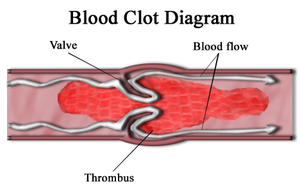
Formation of blood clot (thrombus) in uninjured vessels or thrombic occlusion of a vessel after minor injury. The thrombus if formed of blood elements essentially platelets that develop inside the CVS during LIFE!!

how thrombus occur?


1. Endothelial injury


2.Hypercoaglability

Any alteration of the coagulation pathways that predispose to thrombosis. Eg genetics and sickle cell anemia


3. Abnormal blood flow

Can cause disrupt the laminar flow bringing platelets into contact with endothelium, promote endothelial cell activation, prevent dilution of the activated clotting factors by fresh flowing blood, retard the inflow clotting factors inhibitors--->thrombus formation


Venous thrombus


eg: Deep vein thrombosis (DVT)


Fate of a thrombus

Propagation- Thrombi may accumulate more platelets and fibrin eventually obstructing other critical vessel

Dissolution- Thrombi may be removef by fibriolytic activity

Embolisation-Detach thrombi from vessels may migrate and block small capillaries (emboli)

Organisation and Recanalisation- Thrombi may induce inflammation and fibrosis (organization) and may eventually recanalize

Infection forming mycotic aneurysm and septic embolisation


Embolism

A detached intravascular solid, liquid or gaseous mass that is carried by the blood to a site distant from its point of origin99% of the emboli are dislodged thrombi, hence, the term THROMBOEMBOLISM. Potential consequence of embolism is INFARCTION of the tissue distal to it.


Process of embolism
Types

Thromboembolism
Fat embolism eg after fractures
Air embolism, eg in open carotid injury, angiographic procedures, Caisson’s disease in deep sea divers
Amniotic fluid embolism in abruption placenta of pregnancy


Emboli in brain


Infraction

Area of ischemic necrosis caused by occlusion of either the arterial supply or the venous drainage in a particular tissue.

Example

  • Myocardial Infraction
  • stroke
  • pulmonary infarct following PTE
  • gangrene of limbs due to PVD
  • tortion of testes causing testicular infarction
  • volvulus intussusception causing bowel gangrene

Factors influencing infarction:
  • Nature of blood supply
  • Lungs have dual supply from pulmonary and bronchial arteries
  • Liver has dual supply from hepatic artery and portal vein
  • Kidney, spleen and brain have end arteries with no anastomosis
Rate of occlusion: if slow, can allow for collateral formation, eg in coarctation of aorta

Tissue vulnerability to hypoxia: neural tissue is the most susceptible, dying within 3 to 4 minutes, and myocardium within 30 to 40 min

Oxygen content in the blood: increased risk of infarction in anemic and cyanosed patient

Gross Morphology:
Infarction starts as a poorly defined wedge shaped area, with exudates and hemorrhagic area, which gets more defined by a rim of inflammation after some days.

2 types of infraction (based on their appearance)


Red infarct

Filled with blood. Characterized by coagulation necrosis and erythrocytes from adjacent arteries and veins. Seen with venous occlusions or within loose tissue that allow blood to collect in the infarcted zone of in organs with dual blood supply (lung, liver) or extensive collateral circulation (brain, smallintestine). The latter results because blood flows from the unobstructed vascular
channels into the necrotic area.


Red infact

White infarct

Lacks blood, also called anemic infarct. (Usually has a red zone at periphery because of capillaries at the border of infarct undergo dissolution and blood seeps into the area of necrosis). Occurs with arterial occlusions in solid organs (heart, kidney).


Area of infarct in spleen


Haemorrhage

Escape of blood from the cardiovascular system (extravasation).Discharge of blood from the vascular compartment to the exterior of the body orenclosed within a tissue. Capillary bleeding can occur under conditions of chroniccongestion.


Causes

  • Trauma
  • subcutaneous or intramuscular hemorrhage
  • Septicemia, viremia or toxic conditions
  • widespread petechiae and ecchymoses
  • Coagulation Disorders
  • hemorrhage thrombocytopenia (decreased numbers of platelets)

Origin of hemorrhage

Hemorrhage by rhexis: due to substantial rant or tear present in the blood vessel or heart. Moderate flow of blood out from vascular system


Hemorrhage by diapedisis: due to small defect or rbc passing through the wall in hyperaemia of inflammation


Types of hemorrhage (according to sizes)

1.Petechiae- pin-point foci of haemorrhage up to 1-2 mm in size
2.Purpura- Hemorrhages 3mm. May be associated with diseases which cause petechiae, vascular inflammation or vascular damage. Often scattered on many body surfaces.


Purpura

3. Echymoses- Larger than petechiae and usually blotchy or irregular areas up to >1-2 cm in size often seen with trauma and other problems. (eg: subcutaneous hematomas / bruises)

Echymoses


4. Hematoma- Mass of fibrin and red cells surrounded by vascular connective tissue (supplies nutrients and support for phagocytes (macrophages)


Hematoma


Sources: MBBS Medicine.blogspot.com, circulatory disturbance; docslide.com, circulatory of disturbance; Lisa Miller, General Pathology VPM 152

Ps: This is incomplete article. More references needed


  • Digg
  • Del.icio.us
  • StumbleUpon
  • Reddit
  • RSS
Post Icon

Acid base balance

When it comes to chemistry, it is always about confusion. LOL just kidding :)


***************************************************

Acid base balance

Acid-base balance is critical for maintaining the narrow pH range that is required for various enzyme systems to function optimally in the body.4 Normal blood pH ranges from 7.3-7.4.3 Decreased pH is termed acidemia and is caused by an increase in the concentration of hydrogen ions ([H+]). Increased blood pH is termed alkalemia and is caused by a decrease in the [H+].

The buffer systems that maintain this pH balance are bicarbonate, phosphates, and proteins. Bicarbonate is the most important extracellular buffer (blood), while phosphates and proteins contribute mostly to intracellular acid-base balance.The bicarbonate system is the only buffer measured for the calculation of acid-base status in patients and is represented by the equilibrium equation:

CO2 + H2O <—> H2CO3 <—> H+ + HCO3-


This equation allows one to visualize what effects the addition of carbon dioxide (CO2) or bicarbonate (HCO3-) will have on the buffer system and the blood pH. Addition of CO2 to the system will cause the equation to shift to the right, increasing the [H+] and, therefore, lowering the pH. Addition of HCO3- to the system will cause the equation to shift to the left, lowering the [H+] and increasing the pH.

Another way to conceptualize this information is to simply think of CO2 as an acid and HCO3- as a base. If CO2 is increased it will tend to cause acidemia. If HCO3- is increased, then alkalemia is the expected result.

In addition to buffers, the lungs and kidneys play a major role in acid-base homeostasis.The lungs function in ventilation and they are responsible for regulating the amount of CO2 present in plasma. The kidneys are responsible for controlling the amount of HCO3- in the blood by resorbing or excreting it in the proximal tubule.Abnormalities in acid-base status are classified as to whether the primary abnormality lies with the CO2 concentration or the HCO3-concentration ([HCO3-]). If CO2 is primarily affected, then a respiratory disturbance is present. If HCO3- is primarily affected, then a metabolic disturbance is present.


Acid base disorder

Simple acid-base disorders are those that are confined to one primary alteration in CO2 or HCO3- with or without a compensatory response. There are four simple acid-base disorders: respiratory acidosis, metabolic acidosis, respiratory alkalosis and metabolic alkalosis.


Acidosis is a physiologic condition that tends to increase the concentration of hydrogen ions, which will decrease the pH.This condition can be respiratory or metabolic in origin. An increase in the concentration of CO2, expressed as pCO2, is known as respiratory acidosis. Alternatively, a decrease in the [HCO3-] is known as metabolic acidosis. Both of these situations cause the buffer equation to shift to the right, causing an increased [H+] and a decreased pH.


Alkalosis is a physiologic condition that tends to decrease the [H+], which will increase the pH. Like acidosis, alkalosis can be respiratory or metabolic in origin. A primary condition that results in decreased pCO2 is termed respiratory alkalosis and a primary condition that results in increased [HCO3-] is termed metabolic alkalosis. Both situations shift the bicarbonate equation to the left, resulting in a decreased [H+] and an increased pH.


Acidemia and alkalemia are alterations in the pH of the blood. Acidosis and alkalosis (respiratory or metabolic) are the disorders that will cause these pH alterations. Most cases of simple acidosis or alkalosis, such as those described above, will result in acidemia or alkalemia, respectively.

However, there are compensatory mechanisms that the body will employ in an attempt to maintain a normal pH. In these situations it is possible to have an acidosis or alkalosis disorder and maintain a normal blood pH. There are also mixed acid-base disorders in which opposing primary disorders can effectively cancel each other out and produce a normal pH balance.


Let's a look at one by one

Repiratory Acidosis

Respiratory acidosis is caused by any condition which increases the pCO2 (hypercapnia).While increased production of CO2 (hyperthermia, cardiopulmonary arrest) is a possible cause of hypercapnia, the vast majority of cases are due to impaired removal of CO2 through the lungs. Hypoventilation, ventilation-perfusion mismatch and impaired alveolar gas exchange can all lead to hypercapnia. Therefore, the broad categories of disease which can lead to respiratory acidosis include:

  • respiratory center depression
  • neuromuscular disease
  • restrictive extrapulmonary disease
  • intrinsic pulmonary
  • small airway disease
  • large airway obstruction,
  • increased CO2 production with impaired alveolar ventilation.

Respiratory alkalosis

Respiratory alkalosis is caused by conditions that will decrease the pCO2 (hypocapnia). Hyperventilation will lead to hypocapnia, and it can be caused by hypoxemia, pulmonary disease, direct activation of the respiratory center in the brainstem, overzealous mechanical ventilation, or situations causing pain, fear, or anxiety.


Metabolic acidosis

May result from either an excess of acid or reduced buffering capacity due to a low concentration of bicarbonate. Excess acid may occur due increased production of organic acids or, more rarely, ingestion of acidic compounds.

There are two types of metabolic acidosis. Both are characterized by a decrease in the [HCO3-] but they differ in how that decrease occurs. Secretional metabolic acidosis is caused by a direct loss of bicarbonate-rich fluid such as diarrhea or saliva. Titrational metabolic acidosis is caused by the presence of non-CO2 acids that titrate bicarbonate causing a decreased [HCO3-].

Titration-type metabolic acidosis is the result of increased endogenous or exogenous acids in the plasma. Exogenous acids include ethylene glycol metabolites and salicylate. Endogenous acids include lactic acid, uremic acids, and ketones. It follows then that shock, renal failure, and diabetic ketoacidosis, respectively are common causes of titration-type metabolic acidosis. In cases of hypovolemic shock, perfusion is decreased. Anaerobic metabolism is a consequence of decreased perfusion, so lactic acid accumulates.

In cases of renal failure, uremic acids will accumulate since the kidneys can not effectively excrete them. In cases of undiagnosed or uncontrolled diabetes mellitus, cells cannot utilize glucose due to a lack of insulin. Therefore, the body must metabolize its adipose tissue. Ketones are the byproduct of oxidation of free fatty acids by the liver.


Anion gap

Secretional and titrational metabolic acidosis can be differentiated by their effects on the anion gap (AG). The anion gap is a calculated value based on the principle of electroneutrality which states that the total anions in the body must always be equal to the total cations. We regularly measure the most significant ions: Na+, K+, Cl- and HCO3-. The ions we do not regularly measure are referred to as unmeasured ions.

There are unmeasured cations (Ca+2, Mg+2, and gammaglobulins) and unmeasured anions (albumin, phosphates, sulfates, and organic acids). The unmeasured anions outnumber the unmeasured cations and the difference is the anion gap (Figure 1). The anion gap is easily calculated from the ions we do measure: AG = (Na+ + K+) – (Cl- + HCO3-).4 Unmeasured cations do not undergo significant changes in health or disease and so changes in the anion gap are almost always associated with changes in the unmeasured anions.

Figure 1: Normal anion gap

With secretion-type metabolic acidosis, the anion gap is normal. The body compensates for the increased loss of bicarbonate by a retaining Cl-. As such, as the [HCO3-] decreases, the [Cl-] increases and the anion gap remains normal (Figure 2).


Figure 2: Normal anion gap in secretion acidosis

With titrational metabolic acidosis, the anion gap is increased. Remember that titration type metabolic acidosis is associated with increased levels of exogenous or endogenous acids. Since HCO3- is consumed to buffer these organic acids and there is no effect on the [Cl-], the anion gap increases (Figure 3). The anion gap is therefore very useful in determining a possible etiology for metabolic acidosis that can be confirmed based on history and clinical signs.


Figure 3: Increased anion gap in titration acidosis


Metabolic alkalosis

Causes of metabolic alkalosis include loss of acidic chloride-rich fluids from the body and chronic administration of alkali. In small animal practice, most cases of metabolic alkalosis are caused by vomiting of stomach contents. Abomasal reflux of hydrochloric acid (HCl) into the rumen will cause metabolic alkalosis in ruminants.


Compensation

Compensation is the process whereby the body attempts to restore the normal blood pH during an acid-base disorder. Overcompensation does not occur. As with the primary disorders, compensation can be metabolic or respiratory in origin.

Acid-Base DisturbancePrimary DisturbanceCompensatory ResponseCompensatory Mechanism
Respiratory acidosisIncreased pCO2Increase [HCO3-]Acidic urine
Respiratory alkalosisDecreased pCO2Decreased [HCO3-]Alkaline urine
Metabolic acidosisDecreased [HCO3-]Decrease pCO2Hyperventilation
Metabolic alkalosisIncreased [HCO3-]Increased pCO2Hypoventilation
Compensatory process

Keyword: Respiratory--> compensate at kidney
Metabolic --> compensate at lungs


Diagnosis of acid base disorder

The values that need to be examined to determine if there is an acid-base imbalance are: blood pH, pCO2, [HCO3-], and the anion gap. If any of these values are outside of the reference range, an acid-base abnormality is present. The blood gas analysis reports pH, pCO2, [HCO3-], and pO2. Blood gas analysis is completed on whole blood collected in heparin (green tube), and the sample should be collected anaerobically and processed as soon as possible after collection. The serum chemistry profile reports the anion gap and total CO2 (TCO2). TCO2, not to be confused with pCO2, is another measurement of [HCO3-] and is often used synonomously.

Most simple acid-base disorders are associated with a change in pH. Once acidemia or alkalemia is detected, the next step is to determine if the primary cause is respiratory or metabolic in nature. This requires analysis of the pCO2 and HCO3- (TCO2) values. Changes in the HCO3- value that correspond to the change in pH reflect a primary metabolic disorder while changes in the pCO2 that correspond to the change in pH reflect a primary respiratory disorder. The next step is to determine if there is secondary compensation by the body to try to correct the primary disturbance.


Mixed acid base disorder

A mixed acid-base disorder is one in which two different primary conditions are acting at the same time. Mixed disorders can be a combination of metabolic and respiratory disorders or a combination of different metabolic disorders. The separate processes may have either a neutralizing or additive effect on the pH.

First, there are mixed disorders which have a neutralizing effect on pH. In these cases, the body may appear to be overcompensating because the pH is normal or close to normal. Since the body does not overcompensate, a mixed disorder should be suspected. A classic example of this type of disorder is a vomiting dog who becomes dehydrated. The loss of stomach acid leads to alkalosis while the dehydration and subsequent lactic acid buildup leads to acidosis.

Second, it is possible to have mixed disorders which have an additive effect on pH.2 For example, a respiratory acidosis and metabolic acidosis can occur concurrently in a dog with thoracic trauma that also has lactic acidosis due to shock. In this case the pH would be dangerously low. Mixed disorders that have an additive effective on the pH will always have an abnormal pH.


Effects of acid base disorder

Many of the clinical signs observed in animals with acid-base disturbances are the result of the primary disease process but there are also clinical signs that can develop as a result of the acid-base disturbance itself. Most notably, changes in neural function, cardiac output and concentrations of calcium and potassium may all occur as a direct result of acid-base abnormalities.

Severe acidosis will impair the ability of the brain to regulate its volume, resulting in obtundation and coma. Severe metabolic alkalosis can induce agitation, disorientation, stupor and coma. Cardiac output is also compromised by severe acidosis. Myocardial contractility decreases when the blood pH falls below 7.2 and acidosis may predispose the heart to ventricular arrythmias or ventricular fibrillation.

Serum ionized calcium concentration can be affected by acid-base abnormalities. Acidosis causes displacement of calcium ions from their binding sites on albumin as the binding sites become protonated and an increase in ionized calcium concentration results. Conversely, alkalosis will cause a decrease in ionized calcium concentration and may lead to muscle twitching.

The distribution of potassium ions between the intracellular and the extracellular fluids (and therefore the blood [K+]) may be affected by acid-base disorders as well. As the blood [H+] rises in cases of acidosis, more H+ ions are pumped intracellularly in exchange for K+ ions that are pumped extracellularly (Figure 4). This exchange of ions is necessary to maintain electrical neutrality. The result is a rise in serum [K+]. This situation has only been observed in cases of titration-type metabolic acidosis caused by non-organic (mineral) acids like hydrochloric acid and secretion-type metabolic acidosis.Even though hyperkalemia can develop, it is not likely to occur if renal function and urine output are normal. Clinical signs of hyperkalemia can include muscle weakness and cardiac conduction disturbances.


Figure 4: Exchange oh H+ and K+ in acidosis. Result in hyperkalemia


Just as hyperkalemia can accompany acidosis, hypokalemia can accompany alkalosis.Hydrogen ions moves out of the cell to increase the [H+] extracellularly, so potassium ions move inside the cell to maintain electrical neutrality (Figure 5). Clinical signs of hypokalemia can include muscle weakness, polyuria, polydipsia, impaired urinary concentrating ability, and cardiac arrhythmias.


Figure 5: Exchange of H+ and K+ in alkalosis. May cause hypokalemia


Treatment of acid base disorder

Treatment of respiratory acidosis and respiratory alkalosis is aimed at correcting hypercapnia and hypocapnia, respectively. Therefore, causes of hypoventilation and hyperventilation need to be investigated in order to determine the underlying cause of hypercapnia and hypocapnia, respectively.

Treatment of metabolic alkalosis is aimed at replacing the chloride deficit in cases where vomiting of stomach contents or diuretic administration is the underlying cause.However, potassium and sodium deficits are likely to be present also, so these must be addressed simultaneously. Therefore, the treatment of choice for the correction of metabolic alkalosis in dogs and cats is intravenous administration of a sodium chloride (NaCl) solution with added potassium chloride (KCl). If ongoing gastric losses are present, H2-blocking agents can be useful to decrease gastric acid secretion. In cases where a gastric foreign body is causing vomiting, definitive treatment consists of surgical removal of the foreign body.

Treatment of metabolic acidosis is dependent upon the underlying etiology. Fluid therapy to correct dehydration and electrolyte disturbances, if present, is warranted. However, specific treatments will vary for ethylene glycol intoxication, lactic acidosis, renal failure, and diabetic ketoacidosis.


Ps: Hope this won't be confused no more


Sources: Full article of Acid-Base Balance, an overview; Veterinary Clinical Pathology Clerkship Program

  • Digg
  • Del.icio.us
  • StumbleUpon
  • Reddit
  • RSS
Post Icon

Cardiopulmonary resuscitation (CPR) in animal

Animal CPR (a big LOL)

During anesthetic class, we were taught to perform CPR on emergency anesthesia. This will not happen if the anesthetists are very careful in monitoring the patient throughout surgical period. So, let's dive in!

**********************************************************


CPR in dog


CPR is an emergency technique used to help someone whose heart and/or breathing has stopped. Although somewhat modified, the same techniques used for people – rescue breathing and chest compression – can be used to help treat an animal in distress.

The first lesson to know about CPR is that it doesn't restart a stopped heart. The purpose of CPR, in both humans and animals, is to keep them alive until the heart begins beating on its own or a cardiac defibrillator can be used. In animals, CPR is frequently unsuccessful, even if performed by a trained veterinarian. Even so, attempting CPR will give animal fighting chance.


PCR on cat


At first, we must know the ABC rules!


A-Airway
1.Listen and feel to determine if your pet is breathing/has a pulse.
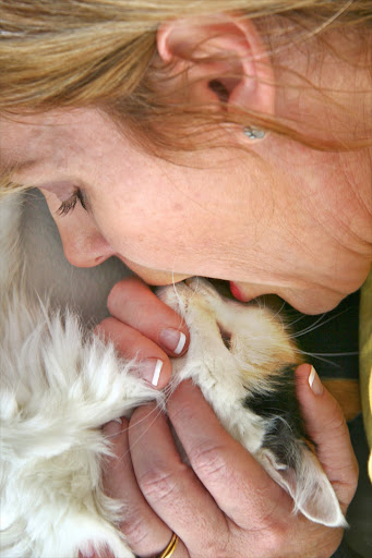
2.Carefully pull the tongue straight out of the animal’s mouth to open the airway
3. Make sure that the neck is reasonably straight; try to bring the head in-line withthe neck.

WARNING: Do not over-straighten the neck in cases where neck/head trauma exists

4. Attempt 2 rescue breaths, by closing the mouth, and performing mouth-to-nose ventilations. If they go in with no problems continue to B-Breathing.

Step 2: Place pet on his right side (legs away from you)...
Opening airway in dog

See above for positioning to give breaths to a cat.

Opening airway in cat

5. If not, reposition the neck and try5. step 3 again.

6.Visibly inspect the airway by looking into the mouth, and down the throat for foreign objects occluding the airway. Unlike human-CPR, rescuers may reach into the airway and remove foreign objects that are visible.

6. If you still can’t breath into the animal,proceed to the Heimlich maneuver.

Heimlich maneuver

Use to remove choking material that cause inability in breathing

1. Turn the animal upside down, with its back against your chest 2. Hug the animal with your fist in your hand, just below the rib-cage ( for cats, just squeeze 1 hand in the same place)


2. With both arms, give 5 sharp thrusts (bear hugs) to the abdomen. Perform each thrust as if it is the one that will expel the object

3. Stop, check to see if the object is visible in the airway, if so, remove it and give 2 mouth-nose rescue breaths. If the breaths do not go in, go back to step 1


Heimlich maneuver


B-Breathing

After achieving a patent airway, one must determine whether the animal is breathing, and whether this breathing is effective:

1. Carefully pull the tongue straight out of the animal’s mouth to open the airway

2. Make sure that the neck is reasonably straight; try to bring the head in-line withthe neck.

3. Breathe at 12 breaths per minute (1 every 5 seconds)With each breath just make the chest rise (do not overinflate, expecially on a small animal)

IMPORTANT: If the breaths do not go in, stop and return to A-Airway!


Proceed to C-Circulation, while continuing breathing support as necessary.


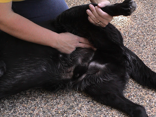
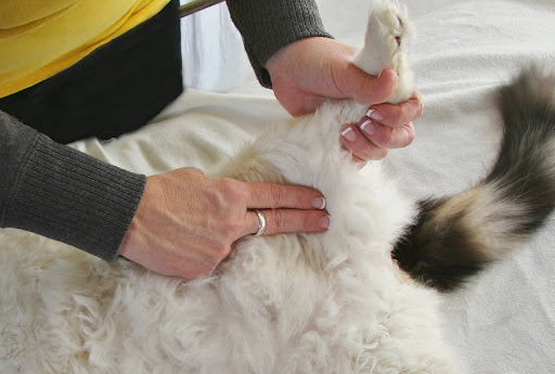
Check the femoral pulse!

Step 3 (Dog): Check pulse at femoral artery (where hind leg meets torso), If no pulse…

Dog

Step 3 (Cat): Check pulse at femoral artery (where hind leg meets torso), if no pulse…

Cat


C- Circulation

This is the final step of CPR and should only be started after the A-airway and B-breathing steps have been completed:

1. Make sure that there are no major (pooling/spurting blood) points of bleeding. Control as necessary by applying pressure with your hand

2. Check for a pulse in the groin (check carefully on a conscious dog or cat!)

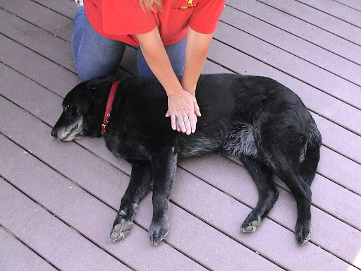
3. Lay the animal on its right side

Step 4 Gently take animal’s left front leg and bend it at the elbow, rotating it at the shoulder.  Where his elbow touches his body is where you place your left hand for compressions.


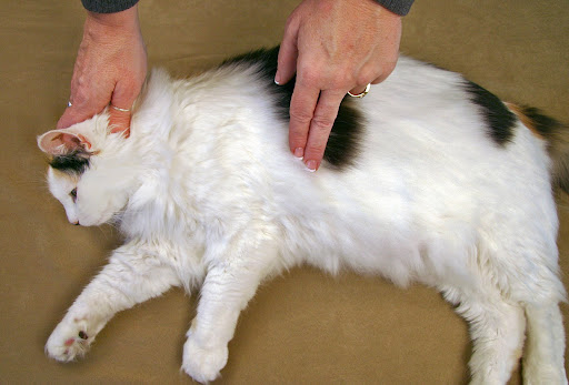
4. Locate your hands where its left elbow touches the chest, approximately the middle of the rib-cage (for cats use 1 hand in asqueezing motion).

Compressions for medium to large dogs: With you left hand, push on the chest 10-15 times (approximately 3 compressions every 2 seconds) and then deliver 2 more breaths. Repeat.  Every 4 cycles, check for a pulse.


Location of heart

Compress the chest 15 times followed by 2 rescue breaths (3 compression every 2 seconds). Deliver 2 more breath. Repeat. Every 4 cycle for a pulse.

Compressions for small cats and dogs:  Use fingertips to compress heart in place of your left hand or place 4 fingers of your left hand under the animal’s chest and compress on the top with left thumb: 5 compressions/1 breath and check for a pulse every 8-10 cycles.

Compression for small cats and dogs: Use fingertips to compress heart in place of your left hand or place 4 fingers of your left hand under the animal’s chest and compress on the top with left thumb: 5 compression/1 breath and check for a pulse every 8-10 cycles.

If there are two people, one breathes and the others compresses at the rate of one breath for every 2-3 compression


After the CPR is successfully performed, quickly send the animal to the nearest vet!


Precaution in CPR

1. Take care not to over inflate the lungs, observe that you ventilate with a normal rise and fall to the chest.

2. Never breathe or compress on an animal that is breathing or has a pulse.

3. When doing compression, realize you must compress 1/4 to 1/3 the width of the chest. You must flex the ribs, press the lungs in order to squeeze the blood out of the heart and release so that it can flow around and back into the other chamber.

4. For barrel-chested breeds, you may position dog on his back and compress chest human-style (hand on top of hand over chest): 15 compressions/2 breaths checking for a pulse every 4 cycles.

For barrel-chested breeds (like Fala and Winnie above) you may position dog on his back and compress chest human-style (hand on top of hand over chest):  15 compressions/2 breaths checking for a pulse every 4 cycles.

Barrel-chested dog


**Perform CPR until you have reached a veterinary hospital. After 20 minutes, however, the chances of reviving an animal are extremely unlikely.


Maybe you can learn something here..


Sources: Sheraton luxuries; CPR guide, CPR in dogs;petplace.com, dailywag; Martha steward.com; Pet first aid

  • Digg
  • Del.icio.us
  • StumbleUpon
  • Reddit
  • RSS