Powered by Blogger.
RSS

Welcome

I created this blog as an instrument of what I have encountered in the world of veterinary medicine as a proud vet student. Comments and suggestions are welcome here at;

sweet_daffodil90@yahoo.co.uk

Regards,
Aina Meducci 2012

Disclaimer

The following blog posts is not genuinely from my research but through readings and citation from trusted website. I do not own any of the copyright and therefore you may use it at your own risk

SINCE I AM NOT A VETERINARIAN YET, THEREFORE I CAN'T CONSULT ANY MEDICAL ADVICE TO YOU AND YOUR PETS! EXTREMELY IMPORTANT!.

Happy reading!
Post Icon

Infectious bovine keratoconjunctivitis (pink eye)

This semester takes me on a endless marathon. To get in class on time, with 3 times a week presentation and followup treatment upon every case we encountered. My weekends are not meant to be free.Not to mentioned the subjects I need to head over heel with and struggling to carry out my duty as in educational and social activities.

Owh, I really need a break to catch up. Didn't have much time to write my blog


Tonight (11.16 PM) we will come into one of my case studies- Moraxella bovis (pink eye)


*******************************************************


Pink eye in cattle


Pinkeye (infectious bovine keratoconjunctivitis or New Forest eye) is a common infectious disease affecting the eyes of cattle. The name describes the redness and inflammation of the lining of the eyelid and eyeball. Although pinkeye is non-fatal, it has a marked economic impact on the cattle industry. It is known to occur at all seasons of the year and in all breeds of cattle.

It is a highly contagious disease, causing inflammation of the cornea (the clear outer layer) and conjunctiva (the pink membrane lining the eyelids) of the eye. It will also cause ulceration, which looks like a hole or depression in the cornea. The incidence of pinkeye increases in spring, peaks in the summer, and decreases in the fall. Pinkeye results in mild to severe disease and, in approximately 2 percent of the cases, will cause blindness.


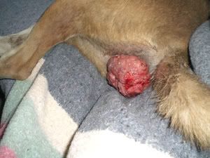
Infected cattle with severe discharge and face flies


Etiology and transmission

Moraxella bovis is the primary infectious agent initiating pinkeye. Other microorganisms initiating pinkeye include Chlamydia, Mycoplasma, and Acholeplasma, or viruses such as the Infectious Bovine Rhinotracheitis (IBR) virus, which can either add to the severity of the disease process or may serve as predisposing factors permitting a secondary infection with M. bovis.Other factors instrumental in causing eye irritation, thereby allowing for invasion of M. bovis and subsequent disease, are excessive ultraviolet light (sunlight), the face fly (Musca autumnalis), the house fly (Musca domestica), the stable fly (Stomoxys calcitrans), plant material, and dust.


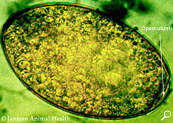
Moraxella bovis is a Gram-negative, aerobic, oxidase-positive diplococcus


Ultraviolet (UV) light is especially a problem for cattle lacking pigmentation around the eye. Lack of pigmentation allows increased UV radiation to sensitize the eye, resulting in inflammation and subsequent infection.Flies not only serve as irritants as they feed on secretions from the eye, but they also serve as a means of transmitting M. bovis from infected to non-infected animals. Face flies can remain infected with M. bovis up to three days following feeding on infected material. Under experimental conditions, disease transmission is uncommon without the presence of face flies and is common with flies present.


Transmission of M. bovis occurs through direct contact, flies, and in-animate objects. The organism is located in the eyes and nasal cavities of infected cattle. Infected secretions from these areas are a source of infection for other cattle. Infected, asymptomatic (no symptoms) cattle may serve as carriers, and will harbor M. bovis in their nasal cavities for a period that may exceed one year. These carrier animals allow for the persistence of pinkeye at a particular site from year to year. Ultraviolet radiation, face flies, growing plants, and pollen production are at their peak in the summer and fall, and account for the high incidence of pinkeye during this period. Weaning distress, increased concentration of cattle, increased exposure to other infectious agents (IBR virus, Mycoplasma, etc.), and hay feeding often are contributing factors to increased disease incidence in late fall, winter, and early spring.


Clinical signs

There are four stages of pinkeye. The disease may resolve at any of these stages while, without treatment, the most severe cases will progress through all four stages.


Stage I: Cattle have excessive tearing and increased sensitivity to light. They will blink frequently and there is redness along the eyelids. Cattle will often seek shade, which will decrease their grazing time. Pain associated with pinkeye also decreases their feed intake. Stage I will progress to a small ulcer in the center of the cornea which appears as a small white spot. The cornea develops a slightly cloudy grey appearance due to inflammation. One or both eyes may be affected.

stage1.jpg


Stage II: The clinical signs described in Stage I continue, but the ulcer spreads across the cornea. As more inflammation occurs, the cornea becomes increasingly cloudy. At this point, some of the dark color of the iris can still be seen. Blood vessels from the outside portion of the cornea begin to grow across the cornea to help with healing. These blood vessels make the cornea appear pink, which is how the disease received its name.


stage2.jpg


Stage III: The ulcer covers most of the cornea and the inflammation continues to spread into the inner parts of the eye. When this occurs, the inside of the eye fills with fibrin, which is a pus-like substance that gives the eye a yellow appearance versus the typical brown appearance.

stage3.jpg


Stage IV: The ulcer extends completely through the cornea, and the iris may protrude through the ulcer. The iris will become stuck in the cornea even after healing. This may lead to glaucoma or persistent swelling of the eye. This eye will be partially or completely blind. The eye may go on to completely rupture, and will develop a shrunken appearance or enlarge if glaucoma (increased eye pressure) is present. This eye will be permanently blind.


stage4.jpg


Inactive scar: Once healing occurs (except Stage IV) the blood vessels will recede, but the eye may continue to be a cloudy blue color. The blue appearance may eventually resolve and the eye appears clear again. In other cases, depending on the severity of the disease, a white scar may be present even after full resolution of the disease.

scar.jpg


Diagnosis

In all species, presumptive diagnosis is based on ocular signs and concurrent systemic disease. It is important to distinguish that the lesions are not due to foreign bodies or parasites. In IBR, upper respiratory signs and conjunctivitis predominate, while keratitis accompanied by ulceration is rare. In bovine malignant catarrhal fever, respiratory signs are prominent with primary uveitis and associated keratitis.

Microbial culture may be beneficial in confirming the causative organisms. Chlamydophila and Mycoplasma spp require special media; the diagnostic laboratory should be consulted prior to sample collection. Cytologic evaluation of stained slides prepared from conjunctival scrapings of sheep and goats may reveal Chlamydophila or Mycoplasma organisms. However, the intracytoplasmic inclusion bodies can be difficult to recognize. PCR analysis can be used to detect Chlamydophila and Mycoplasma spp .


Treatment

Early treatment of cattle with pinkeye is important, not only for a successful outcome of the individual animal affected, but also to stop the shedding of the bacteria to decrease the risk of transmission to other cattle.


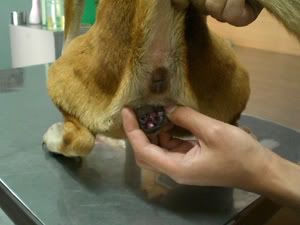
Stage I: Long-acting tetracyclines (Biomycin 200®, LA200®, or their generic equivalents) are effective at this stage of infection. The recommended dose is 4.5 cc per 100 pounds of body weight subcutaneously (SQ). A second injection given 48 to 72 hours later may increase the percentage of cattle that responds to treatment. Another option is to inject penicillin and dexamethasone into the bulbar conjunctiva. The bulbar conjunctiva is the thin membrane that covers the white portion (or sclera) of the eye. If the injection is performed correctly, the conjunctiva will swell and a bulge should be seen in this area. A veterinarian, or someone who has been specifically trained by a veterinarian, should perform this procedure. Injections placed in the wrong area are ineffective in treating pinkeye and could damage the eye.


Stage II: Both tetracycline and a bulbar conjunctival injection are administered at the above dosages

injection1.jpg

Bulbar conjunctival injection


Stage III: Tetracycline and a bulbar conjunctival injection are administered in conjunction with either an eye patch, suturing the third eyelid over the eye, or suturing the eyelids shut. This makes the eye more comfortable, reducing further irritation, and, therefore, reducing tearing and shedding of the bacteria. Suturing the third eyelid over the eye and suturing the eyelid shut also have the advantage of supporting a fragile cornea to help prevent corneal rupture. Again, this procedure should be done by a veterinarian or someone who has been adequately trained.


Stage IV: Same treatment as Stage III

** In my case, I only use terramycin spray


Prevention

Fly control - continues to be necessary due to isolated areas in with a significant face fly population. Insecticide fly tags, sprays, charged backrubbers, and dusts bags are products that can provide chemical control. Manure, weed, and brush management are necessary for total fly control.

Grass, weed, and brush control - Grazing management, brush beating, mowing, and spraying, minimize pollen and mechanical irritation.

Hay and/or feed bunk management - lower overhead hay feeders, spread hay out, do not feed hay containing mature seed heads or cheat grass in overhead feeders or in round bales, increase bunk space to decrease direct contact.Ultraviolet light (sun light) - breed for eyelid pigmentation, introduce Brahman influence into the herd, provide shade or tree rows with ample room to prevent overcrowding.

Disease management – provide proper immunization against viral diseases (IBR and BVD), isolate infected animals, and decrease environmental and nutritional distress.

Ultraviolet light (sun light) - breed for eyelid pigmentation, introduce Brahman influence into the herd, provide shade or tree rows with ample room to prevent overcrowding.

Vaccination – Commercial and autogenous pinkeye vaccines are available.


Sources: Pinkeye in beef cattle:Virginia coorperative extension, Pink eye in cattle Okhlahoma cooperative extension service, The Merck's veterinary manual 10th ed


  • Digg
  • Del.icio.us
  • StumbleUpon
  • Reddit
  • RSS
Post Icon

Canine transmissible venereal tumor (CTVT)

I have came across this topic in class few days ago.

****************************************************
Asha - low reso.jpg

A dog suffering from TVT


Introduction and etiology

Canine transmissible venereal tumor (CTVT), also called transmissible venereal tumor (TVT), Canine transmissible venereal sarcoma (CTVS), Sticker tumor and infectious sarcoma is a histiocytic tumor of the dog and other canids that mainly affects the external genitalia, and is transmitted from animal to animal during copulation or social behavior such as sniffing and licking. It is one of only three known transmissible cancers; another is Devil facial tumor disease, a cancer which occurs in Tasmanian devils. Females are slightly more prone to tumor development than males. It occurs more commonly in young dogs. TVT may also can be transmitted from the infected bitch to puppies


Sniffing (or licking) of healthy intact dog may predispose the transmission of the TVT fromthe infected dog during sexual or social behaviour


TVT are cauliflower-like, pedunculated, and nodular, papillary, or multilobulated in appearance. They range in size from a small nodule (5 mm) to a large mass (>10 cm) that is firm, though friable. The surface is often ulcerated and inflamed and bleeds easily. TVT may be solitary or multiple and are almost always located on the genitalia. They may be transplanted to adjacent skin and oral, nasal, or conjunctival mucosae. The tumor may arise deep within the prepuce or vagina and be difficult to see during cursory examination. This may lead to misdiagnosis if genital bleeding is incorrectly assumed to be hematuria. The tumor is transplanted from site to site and dog to dog by direct contact with the mass. Initially, TVT grow rapidly. Metastasis is uncommon (5%). When metastasis occurs, it is usually to the regional lymph nodes, but kidney, spleen, eye, brain, pituitary, skin and subcutis, mesenteric lymph nodes, and peritoneum may also be sites.


Lesions of TVT on the penis of an infected dog

Up until this moment, there is no exact etiology of how the tumor can be transmitted to the dog. It was believed that the virus is the media that transmits the tumor genes, but there is no clear cut explanation of how this can occur.


Signs and symptom

The infected dogs/pups may experience

  • A visible mass on the penis or vagina (note the external genitalia)
  • Blood dripping from the penis or vagina
  • Frequent licking of the genetalia
  • Similar masses in the mouth or nose (tumors can spread to these sites from direct contact)


Spreading of TVT to the head


Diagnosis

History and physical exam. It is important to know the animal's sexual history. Physical exam should reveal a reddened, possibly bleeding, lobulated tumor. This may not be apparent without careful examination, as it may be covered by the sheath of the penis or the vulva.

Fine needle aspirate. Aspiration of cells from the mass using a needle and syringe usually yields a diagnosis. The cells are examined microscopically to characterize the tumor type.

Fine-needle aspiration reveals a moderately cellular sample of discrete round cells. Anisocytosis (irregular size of cell) and anisokaryosis (irregular size of nucleoli) may be present but usually are not prominent. A few cells may be binucleate. Individual cells usually have a round to reniform nucleus with inapparent nucleoli. Folds or clefts in the nuclear membrane sometimes produce a unique appearance called a "butt cell" (wherein the nucleus resembles buttocks). The cytoplasm is typically light blue and lacks both vacuoles and granules. Mitotic figures are common. Inflammatory cells, especially lymphocytes and plasma cells, are often dispersed among the tumor cells, especially during regression of the neoplasm.


Histiocytoma, Wright-Leishman stain. Neoplastic cells have a round to oval nucleus and light blue cytoplasm that lacks vacuoles and granules. A mitosis (bottom center) and "butt cell" (upper left) also are present.


Complete blood count and biochemical profile. These are routine blood tests that should be performed prior to medical treatment of the tumor. They should be normal in animals with TVT.


Treatment

Chemotherapy is very effective for TVT, but surgery alone often leads to recurrence. Surgery may be difficult due to the location of these tumors. The prognosis for complete remission with chemotherapy is excellent.The most common chemotherapy agents used for TVT are vincristine, vinblastine, and doxorubicin.Radiation therapy may be effective when chemotherapy does not work.

Daeng 1st treatment

Before treatment

Daeng after just a month

Effect of treatment after 1 month


Home care and Prevention

Monitor for response to therapy and prevent self-trauma by putting an Elizabethan collar on per dogs. Spay or neuter the dogs and do not allow exposure to unknown dogs.


Sources: TVT, Wikipedia.org, TVT, petplace.com, Merck's veterianary manual, Canine round cell tumor, Vet.clinical pathology clerckship program


  • Digg
  • Del.icio.us
  • StumbleUpon
  • Reddit
  • RSS
Post Icon

Histopathology: Tissue slide preparation


Hey guys, I have just started sem 6 this week and it was very tiring week I guess. Nevermind about that, today I would like to repost about preparation of histology slide because initially it was in my blog's draft due to limited information and I have no time to review. But few days ago, I was assigned to find a pathology case in VSD and they gave me an opportunity to prepare the histopath slide of myself!. Here's the real photos


************************************************************

Histopahology tissue slide preparation

1. Organ sample


After post mortem, the organ sample is taken in put in the bottle with 10% buffered formalin. If the size of organ is too big (eg Bull), cut of approximately 1.5 cm square area. Cut only the area of the organs showing the abnormalities (eg haemorrhage, ulceration)

The organ sample in the formalin is left overnight (at least-to proceed next process) to allow full absorption of the formalin. The function of formalin is to preserve the sample and kills the bacteria causing autolysis. The pH must be 7.2 (thats why the formalin is added with buffered)


2. Trimming of tissues


Trimming of organ


Tissue cassette

When the organ is taken out from the formalin, it goes to the second step that is trimming of organ sample into small pieces. The organ is cut in various shapes, such as triangular or square shape (depends of pathologist's preference) and next is place in the tissue cassette. The best suggestion is to put abnormal piece of organ such as kidney together with the normal piece of kidney in the same tissue cassette to allow the pathologist to distinguish the normal cell and abnormal cell of the particular organ.

4. Tissue processing- Dehydration


Modern Histokinate


After the tissue is fixed into the cassette, next it must be place in the histokinate for dehydration process. The tissue is immersed automatically in the low concentration of alcohol to high concentration of alcohol (100%) Once the tissue is dehydrated, next it is immersed in the xylene or chloroform to clear the water and covered with wax to made the tissue easier to cut in the later process. The dehydration process takes about 24 hours.


5. Tissue processing- Embedding with paraffin


Embedding in the paraffin wax


Tissue block


When the tissues is taken out from the histokinate, it will be taken to the embedding machine (I forgot the name!) to start to make paraffin block. The block is made from the melting paraffin wax and it will freeze within about 2 minute within the cassette.


6. Tissue processing- Cutting the sample


Tissue block on the microtome


The tissue block is now can be trim and cut by using microtome. The microtome's function is to cut the tissue block into a very thin piece of film (about 5 micrometer). One must ensure to trim the tissue carefully before taking the film to put in the water bath.

7. Tissue processing- "Fishing" in the water bath


Tissue's film in the water bath

The tissue's film must be immersed in the water bath (45 degree celcius) to remove the wax for staining purposes later. A glass slide covered with 1 drop of albumin is used to "fish" the film from the water bath. The function of the albumin is to adhere the film into the center of the slide.


Fishing the film


8. Tissue processing- Rehydration, staining and dehydration

After the tissue is adhere to the slide, proceed to the rehydration process. Rehydration process is to allow to tissue to gain water and therefore able to pick up the stain (H&E) to be able to see under the light microscope. The process is begins with immersion of the slide into the xylene 1, xylene 2 and xylene 3, 100% alcohol ---> 75% alcohol ---> distilled water ---> hematoxyline--> distilled water ---> 0.5% alcohol acid ---> 0.1% lithium carbonate --> 0.5% eosine--> alcohol concentration from low to high (75%, 90%, 100%,100%,100%) ---> xylene


The staining materials


9. Tissue processing- Mounting


After the slide has been stained, a cover slip must be put on top of the sample. Before that, depex is applied on the sample and mounted with a cover slip. The function of Depex is to fixed the tissue slide so that it is become adhere and therefore it does not remove from the slide. Once the mounting is done, the tissue slide is now ready to be observe under the light microscope.


Label the slide, and histopath complete!


Read it for me please?


Sources: Pathology Unit: Veterinary Service Department Kubang Kerian, Kota Bharu,Kelantan


  • Digg
  • Del.icio.us
  • StumbleUpon
  • Reddit
  • RSS
Post Icon

Fasciola Hepatica (Liver fluke)

Just finished up my field work with few of my colleagues doing some investigation in farms around Bachok, Kelantan. As we had had encountered few cases involving various types of ruminants, I was assigned to present fiver fluke manifestation in ruminants particularly in cattle which are commonly found there.

*********************************************************


F. Hepatica


Fasciola hepatica is the species of parasitic flatworms that infects the liver of various mammals, including humans. The disease caused by the organism is called Fascioliasis or Fasciolosis. It is a trematode species of phylum platyhelminthes. F. hepatica is distributed worldwide, and causes great economic losses in sheep and cattle. It has been known as an important parasite of sheep and cattle for hundreds of years.

Susceptible animal hosts for Fasciola species include:Domestic animals: cattle, sheep, pigs, buffaloes and donkeys;Other domestic animals: horses, goats, dromedaries, camels and llamas; (hares, rabbits and rodents).


F. Hepatica in the liver of infected animal


Morphology

Fasciola hepatica is one of the largest flukes of the world, reaching a length of 30mm and a width of 13mm. It is leaf shaped, pointed posteriorly and wide anteriorly, although the shape varies somewhat. The oral sucker is small but powerful and is located at the end of a cone-shaped projection at the anterior end. The acetabulum is larger than the oral sucker and is anterior. The tegument is covered with large, and scalelike spines.

The intestinal ceca are highly dendritic and extend to near the posterior end of the body. The testes are large and greatly branched, arranged in tandem behind the ovary. The smaller, dendritic ovary lies on the right side, coiling between the ovary and the preacetabular cirrus pouch. Vitelline follicles are extensive, filling most of the lateral body and becoming confluent behind the testes.


F. Hepatica


Life cycle


Life cycle of F.hepatica


The shape of the parasite looks like a leaf, with one end tapering. Each individual parasite posseses ovaries and testes, making them hermaphroditic and able to produce fertilized eggs. The organism attaches to bile ducts with two powerful suckers on each end of its body. The bile ducts are where the mature flukes and release eggs. The eggs are passed out in the host's feces. At temperatures over 10 degrees Celcius, the eggs hatch and release miracidiae within two weeks. The hatchlings must find a Lymanaea snail host within 24 hours of hatching or they will die.

Fasciola hepatica egg

F. Hepatica egg under microscope magnification


After identifying a host snail, the hatchling Fasciola heptica burrow in and begin developing into sporocyst. The germinal cells of the sporocyst develop into rediae which migrate to the hepato-pancreas of the snail. The rediae then develop into cercariae which pass through the snail into water. Cercariae encyst apon nearby plant matter for several months, where they can be ingested by a mammal. Once a mammal ingests these metacercariae, the pH levels of the animals stomach allow the Metacercariae to lose their shell. The cercariae freed from their cyst capsules, burrow through the stomach lining and find their way to the liver. The immature flukes feed on the liver tissue for 6 to 8 weeks, causing the symptoms of acute infection. At this stage the flukes move into the bile ducts where they mature and release eggs, completing it's life cycle. Adult females can produce up to 25000 per day.


Lymnea truncatula
Intermediate host for F.Hepatica larval stage


Clinical signs

Fasciolosis ranges in severity from a devastating disease in sheep to an asymptomatic infection in cattle. The course usually is determined by the number of metacercariae ingested over a short period. In sheep, acute fasciolosis occurs seasonally and is manifest by a distended, painful abdomen; anemia; and sudden death. Deaths can occur within 6 wk of infection. The acute syndrome must be differentiated from “black disease.”

In subacute disease, survival is longer (7-10 wk), even in cases with significant hepatic damage, but deaths occur due to hemorrhage and anemia. Chronic fasciolosis is seen in all seasons; signs include anemia, unthriftiness, submandibular edema, and reduced milk secretion, but even heavily infected cattle may show no clinical signs. Heavy chronic infection is fatal in sheep.Sheep do not appear to develop resistance to infection, and chronic liver damage is cumulative over several years. In cattle, there is evidence of reduced susceptibility after fibrosis of liver tissues and calcification of bile ducts.


Bottle jaw (Submandibular edema) in the infected cattle

In human**

The acute phase of infection in humans is characterized by the migration of immature worms through the liver; hemorrhage and inflammation of the liver can be severe, including fever, abdominal pain, respiratory disturbances and skin rashes. The chronic phase starts when the worms move into the bile ducts; symptoms are nonspecific and usually mild. However, progressive inflammation, leads to fibrosis and thickening of the walls of the ducts and gallbladder that eventually may result in blockage due to the parasites, or their debris. In this case, abdomen pain is a characteristic symptom. Chronic infections can lead to biliary cirrhosis with scarring and fibrosis of the liver and growth deficiencies.


Lesions

Immature, wandering flukes destroy liver tissue and cause hemorrhage. In acute fasciolosis, damage is extensive; the liver is enlarged and friable with fibrinous deposits on the capsule. Migratory tracts can be seen, and the surface has an uneven appearance. In chronic cases, cirrhosis develops. Mature flukes damage the bile ducts, which become enlarged, or even cystic, and have thickened, fibrosed walls. In cattle, the duct walls become greatly thickened and often calcified. Flukes may be found in aberrant sites, eg, lungs. Mixed infections with Fasciola magna can be seen in cattle.


Tissue destruction by wandering flukes may create a microenvironment favorable to activation of clostridial spores. (C.Noyvi type B- Black disease)


Little damage is done by juveniles penetrating the intestinal wall and the capsule surrounding the liver but much necrosis results from migration of flukes through the liver parenchyma. During this time, they feed on liver cells and blood. Anemia sometimes results from heavy infections. Worms in bile ducts cause inflammation and edema, which in turn stimulate production of fibrous tissue in the walls of these ducts. Thus thickened, the ducts can handle less bile and are less responsive to needs of the liver. Back pressure causes atrophy of liver parenchyma, with concomitant cirrhosis and possibly jaundice. In heavy infections the gall bladder is damaged, and walls of the bile ducts are eroded completely.


Fibrous liver tissue


Diagnosis

The oval, operculated, golden brown eggs, 130-150 × 65-90 µm, must be distinguished from those of paramphistomes (rumen flukes), which are larger and clear. Eggs of F hepatica cannot be demonstrated in feces during acute fasciolosis. In subacute or chronic disease in cattle, the number varies from day to day, and repeated fecal examination may be required.

Diagnosis can be aided by an ELISA (commercially available in Europe) that enables diagnosis ~2-3 wk after infection and well before the prepatent period. Plasma concentrations of γ-glutamyltransferase, which are increased with bile duct damage, are also helpful during the late maturation period when flukes are in the bile ducts. At necropsy, the nature of the liver damage is diagnostic. Adult flukes are readily seen in the bile ducts, and immature stages may be squeezed or teased from the cut surface.


Treatment

Drug of choice: Bithionol or Triclabendazole

Note: Praziquantel, a common drug for treating trematode infections is not very effective against Fasciola hepatica.


Prevention and control

Control measures for F hepatica ideally should involve removal of flukes in affected animals, reduction of the intermediate host snail population, and prevention of livestock access to snail-infested pasture. In practice, only the first of these is used in most cases.

While molluscicides can be used to reduce lymnaeid snail populations, those that are available all have drawbacks that restrict their use. Copper sulfate, if applied before the snail population multiplies each year, is effective but toxic to sheep, which must be kept off treated pasture for 6 week after application. Prevention of livestock access to snail-infested pasture is frequently impractical because of the size of the areas involved and the consequent expense of erecting adequate fencing.


Sources: Merck Veterinary Manual, Wikipedia: F. Hepatica, F.Hepatica, IVD research.com


  • Digg
  • Del.icio.us
  • StumbleUpon
  • Reddit
  • RSS