Powered by Blogger.
RSS

Welcome

I created this blog as an instrument of what I have encountered in the world of veterinary medicine as a proud vet student. Comments and suggestions are welcome here at;

sweet_daffodil90@yahoo.co.uk

Regards,
Aina Meducci 2012

Disclaimer

The following blog posts is not genuinely from my research but through readings and citation from trusted website. I do not own any of the copyright and therefore you may use it at your own risk

SINCE I AM NOT A VETERINARIAN YET, THEREFORE I CAN'T CONSULT ANY MEDICAL ADVICE TO YOU AND YOUR PETS! EXTREMELY IMPORTANT!.

Happy reading!
Post Icon

Pathology: Types of necrosis

It's been a while since my last post. I was very busy last week so I dont have much time to update this blog. I'm just coming back from Ipoh, after attending Veterinary Association Malaysia congress and it was awesome. I can imagine myself standing there and present my research paper. Haha. Ok2, back to business!

Just started my pathology class and Prof Imad taught the first things about tissue injury and as well as necrosis.

******************************************************

Types of necrosis


The death of cells in a living tissue is called Necrosis. Necrosis is commonly referred to as a bedsore. The degradation of the tissue starts with swelling within the cell and ultimately an interruption of the membranes of the cell. The organelles begin to breakdown and this spreads and causes an inflammation. Necrosis can be caused due to an injury or infection of some kind. If the infection is not tended to correctly, the chances of necrosis starting is more likely. Necrosis is more severe than other cell deaths in infections because once those cells die they can release chemicals that are harmful or will damage other cells.


example of necrosis: death tissue in LIVING cell


According to Prof Imad, there are 4 major types of necrosis namely; coagulative, liquefaction, caseous, and fat necrosis.


1. Coagulative necrosis


Coagulative: Caused by ischemia. Ischemia results in decreased ATP, increased cytosolic Ca++, and free radical formation, which each eventually cause membrane damage.

  • Decreased ATP results in increased anaerobic glycolysis, accumulation of lactic acid, and therefore decreased intracellular pH.
  • Decreased ATP causes decreased action of Na+ / K+ pumps in the cell membranes, leading to increased Na+ and water within the cell (cell swelling).
  • Other changes: Ribosomal detachment from endoplasmic reticulum; blebs on cell membranes, swelling of endoplasmic reticulum and mitochondria.
  • Up to here, the changes are reversible if oxygenation is restored by reversing the ischemia. If the ischemia continues, necrosis results, causing the cytoplasm to become eosinophilic, the nuclei to lyse or fragment or become pyknotic (hyperchromatic and shrunken). In the early stages of necrosis, the cells remain for several days as ghosts of their former selves, allowing one to still identify them and the tissue (in contrast to the other types of necrosis). The cellular reaction is polys, followed by a granulation tissue responses.


Coagulative necrosis

Characterised by preservation of tissue achitecture, increased cytoplasmic eosinophilia and nuclear changes. The cell may look pyknosis (chromatin clumping and shrinking: increased eosinophilia), karyorrhexis (fragmentation of chromatin), karyolysis (fading of chromatin material) and dissapearance of stainable nuclei


2. Liquefactive necrosis


Usually caused by focal bacterial infections, because they can attract polymorphonuclear leukocytes. The enzymes in the polys are released to fight the bacteria, but also dissolve the tissues nearby, causing an accumulation of pus, effectively liquefying the tissue (hence, the term liquefactive).

The result of hydrolysis. When the cells die, they are rapidly destroyed by lysosomal enzymes, either their own or those from neutrophilic leukocytes (i.e., bacterial infections), or clostridia or snake poison. Acid and lye burns represent the extreme of liquefaction. Also, if both neurons and glia are killed, dead brain liquefies rapidly.

Liquefactive necrosis that is caused by neurophilic leukocytes is called PUS. The term may also be used for an effusion that is full of dead neutrophils. Because of the extensive protein hydrolysis (i.e., more total molecules), there's a tremendous increase in the osmotic pull. This explains the familiar high-pressure in a ripened pimple -- and deep in the brain, for example, this is even more serious.

NOTE: The truism that "brain liquefies" is a common source of misunderstanding. Brain deprived of its oxygen for a few moments will suffer neuronal damage but not necrosis. Brain deprived of blood flow for a few moments longer will lose neuronal structure but not glia, and remain solid. The same is true of diseases in which neurons die off one at a time (i.e., Alzheimer's disease causes the brain to shrivel but not to liquefy). Only if the glia are killed does the brain melt away, and then only after several days.


Liquefactive necrosis in the brain


liquefactive necrosis of the brain demonstrates many macrophages at the right which are cleaning up the necrotic cellular debris.


3. Caseous necrosis


All of the cells in an area die, the tissue architecture is obliterated, and they turn into a crumbly ("friable"), readily-aerosolized powder.

A distinct form of coagulative necrosis seen in mycobacterial infections (e.g., tuberculosis), or in tumor necrosis, in which the coagulated tissue no longer resembles the cells, but is in chunks of unrecognizable debris. Usually there is a giant cell and granulomatous reaction, sometimes with polys, making the appearance distinctive.


Gross appearance of caseous necrosis in a hilar lymph node infected with tuberculosis. The node has a cheesy tan to white appearance. Caseous necrosis is really just a combination of coagulative and liquefactive necrosis that is most characteristic of granulomatous inflammation.


Found in granulomatous inflammation; manifestation of partial immunity of interaction T-Lymphocyte, macrophage, and cytokines associated with tubercolosis. Architecture is not preserved but tissue is not liquefied. Grossly soft and cheeselike. Histologically amorphous and acidophilic.


4. Fat necrosis


A term for necrosis in fat, caused either by release of pancreatic enzymes from pancreas or gut (enzymic fat necrosis) or by trauma to fat, either by a physical blow or by surgery (traumatic fat necrosis). The effect of the enzymes (lipases) is to release free fatty acids, which then can combine with calcium to produce detergents (soapy deposits in the tissues). Histologically, one sees shadowy outlines of fat cells (like coagulative necrosis), but with Ca++ deposits, foam cells, and a surrounding inflammatory reaction.


This is fat necrosis of the pancreas. Cellular injury to the pancreatic acini leads to release of powerful enzymes which damage fat by the production of soaps, and these appear grossly as the soft, chalky white areas seen here on the cut surfaces.


Fat necrosis adjacent to pancreas is seen here. There are some remaining steatocytes at the left which are not necrotic. The necrotic fat cells at the righthave vague cellular outlines, have lost their peripheral nuclei, and their cytoplasm has become a pink amorphous mass of necrotic material.


Other types of necrosis


5. Gangrenous necrosis

Due to cut off blood supply to lower extremities or bowel due to vascular occlusion.

GANGRENE ("gangrenous necrosis") is not a separate kind of necrosis at all, but a term for necrosis that is advanced and visible grossly. If there's mostly coagulation necrosis, (i.e., the typical blackening, desiccating foot that dried up before the bacteria could overgrow), we call it DRY GANGRENE. If there's mostly liquefactive necrosis (i.e., the typical foul-smelling, oozing foot infected with several different kinds of bacteria), or if it's in a wet body cavity, we call it WET GANGRENE.

Occur to most diabetic patients


Gangrenous necrosis at lower limb (dry gangrene)


wet gangrene (bacterial rich)


6. Fibrinoid necrosis

Due to the deposition of fibrin-like material on arterial walls due to immune-mediated vasculitis.


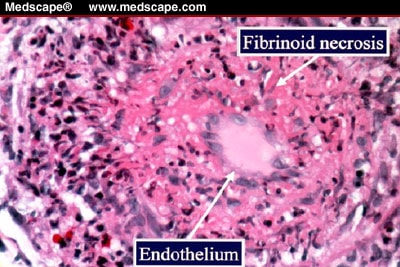
Fibroid necrosis: smudgy pink material in vascular walla with or without necrosis


Sources: USMLE wiki: Cell pathology; cell injury and death Ed Friedlander M.D pathologist; cell injury and necrosis www.uvm.edu; cell injury, library.med.utah.edu

  • Digg
  • Del.icio.us
  • StumbleUpon
  • Reddit
  • RSS
Post Icon

Types of neurotransmitter

During anesthesiology class, we always talk about how the drug goes into the CNS, thereby produce pharmacological effect on the brain by reducing some of the neurotransmitter receptor in order to function. Then I started to think, animal and humans have a lot of neurotransmitters which play many important roles maintaining body system, and who are they??

Ps: Just a basic physiology nervous system revision

***************************************************

NEUROTRANSMITTER


Neurotransmitters are the chemicals responsible for signal transmission between the individual neurons. Most neurons make two or more neurotransmitters, which are released at different stimulation frequencies. 50 or more neurotransmitters have been identified. It is classified by chemical structure and by function.


Criteria to Classify a Biochemical Substance as Neurotransmitter:

  • Substance must be present in the pre-synaptic nerve terminal and packaged into synaptic vesicles ·
  • The substance must be released from the nerve terminal upon arrival of action potential or depolarization of presynaptic membrane ·
  • Specific receptors must be present on the post synaptic membrane for the substance


Life cycle of a neurotransmitter


1.Synthesis of the transmitter
2.Packaging and storage in Synaptic vesicles
3.If necessary, transport from the site of synthesis to the site of release from the nerve terminal 4.Release in response to an action potential
5. Binding to postsynaptic receptor proteins
6.Termination of action by diffusion, destruction, or reuptake into cells.


Chemical classification of neurotransmitter

1. Acetylcholine
2. Biogenic amine
  • Catecholamine: Dopamine, noreprinephrine (NE), and epinephrine (Epi)
  • Indolamines
  • Serotonin and dopamine

3. Amino acid
  • GABA- Gammbaaminobutyric acid
  • Glycine
  • Aspartate
  • Glutamate

4.Neuropeptidase
  • Substance P
  • Endorphines
  • Somatostatin, gastrin, cholecystokinin, oxytoxin, vasopressin, leutanizing hormone releasing hormone

5. Purines
  • Adenosine
  • ATP

6. Gases and Lipid
  • Nitric oxide
  • Carbon monoxide
  • Cannabinoids


Functional classification of neurotransmitter

1. Excitatory neurotransmitter
2.Inhibitory neurotransmitter


Below are some examples of neurotransmitter

Acetycholine

Acetylcholine (Ach) was the first neurotransmitter to be identified It is the most abundant neurotransmitter in the brain. Released at neuromuscular junctions and some ANS neurons Synthesized by enzyme choline acetyltransferaseDegraded by the enzyme acetylcholinesterase (AChE)


Formation and degradation of Ach


Acetylcholine has many functions: It is responsible for much of the stimulation of muscles, including the muscles of the gastro-intestinal system. It is also found in sensory neurons and in the autonomic nervous system (parasympathetic), and has a part in scheduling REM (dream) sleep.


Catecholamine

Catecholamines-Dopamine, norepinephrine (NE), and epinephrine are synthesized from Tyrosines involved in reward-pleasure and learning. Dopamine is the principle neurotransmitter involved in Addiction pathway

1. Norephrineprine

Norepinephrine is strongly associated with bringing nervous systems into "high alert." It is prevalent in the sympathetic nervous system, and it increases our heart rate and our blood pressure. Adrenal glands release it into the blood stream, along with its close relative epinephrine (adrenalin). It is also important for forming memories.Stress tends to deplete our store of adrenalin, while exercise tends to increase it. Amphetamines ("speed") work by causing the release of norepinephrine, as well as other neurotransmitters called dopamine and seratonin.


2. Dopamine

Dopamine has many functions in the brain, including important roles in behavior and cognition, voluntary movement, motivation, punishment and reward, inhibition of prolactin production (involved in lactation and sexual gratification), sleep, mood, attention, working memory, and learning.


3. Serotonin

Serotonin is an inhibitory neurotransmitter that has been found to be intimately involved in emotion and mood. Too little serotonin has been shown to lead to depression, problems with anger control, obsessive-compulsive disorder, and suicide. Too little also leads to an increased appetite for carbohydrates (starchy foods) and trouble sleeping, which are also associated with depression and other emotional disorders. It has also been tied to migraines, irritable bowel syndrome, and fibromyalgia.

Broadly distributed in the brain, derived from Tryptophan involved in sleep, dreaming, hunger and arousal. Play roles in emotional behaviors and the biological clock. Depletion of serotonin in brain leads to depression.


4. GABA

GABA—Gamma-aminobutyric acid is the major inhibitory neurotransmitter in CNS synthesized from decarboxylation of Glutamate involved in regulating anxiety may be related to eating or sleep disorders

GABA acts like a brake to the excitatory neurotransmitters that lead to anxiety. People with too little GABA tend to suffer from anxiety disorders, and drugs like Valium work by enhancing the effects of GABA. Lots of other drugs influence GABA receptors, including alcohol and barbituates. If GABA is lacking in certain parts of the brain, epilepsy results.


5. Endorphins, Enkephalins and Substance P
Substance P is the mediator of pain signals. Endorphins and Enkephalins act as natural opiates; reduce pain perception. They also depress physical functions like breathing and may produce physical dependence.

Endorphin is short for "endogenous morphine." It is structurally very similar to the opioids (opium, morphine, heroin, etc.) and has similar functions: Inhibitory, it is involved in pain reduction and pleasure, and the opioid drugs work by attaching to endorphin's receptor sites. It is also the neurotransmitter that allows bears and other animals to hibernate.


6. Endocannabinoids
Endocannabinoids are lipid soluble; synthesized on demand from membrane lipids. Bind with G protein–coupled receptors in the brain. Involved in learning and memory.


7. Glutamate

Glutamate is an excitatory relative of GABA. It is the most common neurotransmitter in the central nervous system - as much as half of all neurons in the brain - and is especially important in regards to memory. Curiously, glutamate is actually toxic to neurons, and an excess will kill them. Sometimes brain damage or a stroke will lead to an excess and end with many more brain cells dying than from the original trauma. ALS, more commonly known as Lou Gehrig's disease, results from excessive glutamate production. Many believe it may also be responsible for quite a variety of diseases of the nervous system, and are looking for ways to minimize its effects


Functional Classification of Neurotransmitters

Neurotransmitter effects may be excitatory (depolarizing) and/or inhibitory (hyperpolarizing). Determined by the receptor type of the postsynaptic neuron.
  • GABA and glycine are usually inhibitory.
  • Glutamate is usually excitatory
  • Acetylcholine-Excitatory at neuromuscular junctions in skeletal muscle

Neurotransmitter Actions

1. Direct action
  • Neurotransmitter binds to channel-linked receptor and opens ion channels
  • Promotes rapid responses
  • Examples: ACh and amino acids

2. Indirect action
  • Neurotransmitter binds to a G protein-linked receptor and acts through an intracellular second messenger
  • Promotes long-lasting effects
  • Examples: biogenic amines, neuropeptides, and dissolved gases

Neurotransmitter Receptors
1. Channel-linked receptors
2. G protein-linked receptors


Channel-Linked (Ionotropic) Receptors

Ligand-gated ion channels. Action is immediate and brief. Excitatory receptors are channels for small cation. Na+ influx contributes most to depolarization. Inhibitory receptors allow Cl– influx or K+ efflux that causes hyperpolarization.


G Protein-Linked (Metabotropic) Receptors

Transmembrane protein complexes. Responses are indirect, slow, complex, and often prolonged and widespread. Examples: muscarinic ACh receptors and those that bind biogenic amines and neuropeptides


Sources: General psychology Dr C George Boeree


  • Digg
  • Del.icio.us
  • StumbleUpon
  • Reddit
  • RSS