Powered by Blogger.
RSS

Welcome

I created this blog as an instrument of what I have encountered in the world of veterinary medicine as a proud vet student. Comments and suggestions are welcome here at;

sweet_daffodil90@yahoo.co.uk

Regards,
Aina Meducci 2012

Disclaimer

The following blog posts is not genuinely from my research but through readings and citation from trusted website. I do not own any of the copyright and therefore you may use it at your own risk

SINCE I AM NOT A VETERINARIAN YET, THEREFORE I CAN'T CONSULT ANY MEDICAL ADVICE TO YOU AND YOUR PETS! EXTREMELY IMPORTANT!.

Happy reading!
Post Icon

Torticollis in rabbits

A brief conversation with 2 of my classmates

Teng: Rabbit itu kena bacterial infections (That rabbit is having bacterial infections)
Me: Apa bacterial infections? (What bacterial infections?)
Luqman: Arnab tu kepala pusing kebawah sebab penyakit bakteria (That rabbit has twisted neck due to bacterial infections)
Me: Torticollis?
Luqman: Hah! thats it.


**********************************************


Torticollis (head tilt) in rabbit

Torticollis, or wryneck, is a stiff neck associated with muscle spasm, classically causing lateral flexion contracture of the cervical spine musculature (a condition in which the head is tilted to one side). The muscles affected are principally those supplied by the spinal accessory nerve. Generally it can affect rabbits, guinea pigs, and other rodents species as well.


Possible causes of torticollis

  • Middle/inner ear infection (otitis media /interna)
  • Stroke (cerebrovascular accidents)
  • Trauma
  • Cancer (neoplasia)
  • Cervical muscle contraction
  • Encephalitozoonosis
  • Cerebral larva migrans
  • Intoxication

Lets go detail of the causes

1. Middle/inner ear infection

An inner ear infection may have started with an outer ear infection, which remained unnoticed and untreated and gradually worked its way into the inner ear, or with a middle ear infection, which resulted from an upper respiratory infection.

Or it may have arisen from bacteria in the nasal cavity or bloodstream. A radiograph of the head may help determine if the middle ears are affected. Some of the bacteria which have been cultured from ear infections are Staphylococcus sp, Pseudomonas aeruginosa, Pasteurella multocida, Bordetella bronchiseptica, Proteus mirabilis, Streptoccus epidermidis, Bacteroides spp. and Escherichia coli.

Bacterial infections that occur secondary to ear mite infestations can sometimes track deep into the rabbit's ear canal, resulting in a nasty bacterial ear canal infection termedotitis externa. Provided that these bacterial organisms remain located only in the outerear canal of the rabbit (i.e. the external ear canal situated on the outermost aspect of the rabbit's ear drum), systemic antibiotics and topical antibiotic ear medications (in addition to the mite killing treatments) will often be enough to resolve the problem.
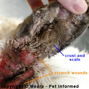
Ear mites in rabbits - the left ear of this rabbit is infested with Psoroptes cuniculi mites.

Mite infection with secondary bacterial infections

Occasionally, however, bacterial organisms will manage to eat their way through the ear drum of ear mite infested rabbits, resulting in a nasty bacterial infection of the middle and inner sanctums of the rabbit's ear (termed the middle ear and inner ear, respectively). These middle-ear and inner-ear bacterial infections (termed, respectively, otitis mediaand otitis interna) are not only very difficult to treat and control but they can result in some serious complications of their own.

Damage to the middle and/or inner ear mechanisms can result in deafness of the ear. Damage to the inner ear mechanisms, in particular the vestibular apparatus of the inner ear (a series of specialized fluid canals located deep within the inner ear, which are responsible for normal balance), can result in the rabbit showing severe signs of incoordination and neurological dysfunction. The rabbit will appear off-balance, falling frequently, often with its head twisted around Bacterial infections that occur secondary to ear mite infestations can sometimes track deep into the rabbit's ear canal, resulting in a nasty bacterial ear canal infection termedotitis externa. Provided that these bacterial organisms remain located only in the outerear canal of the rabbit


2. Stroke

Stroke is usually suspected on the basis of physical signs. Imaging to diagnose this problem is available to humans but difficult to arrange for companion rabbits. As in humans, acerbrovascular accident can kill, but if it does not, then the rabbit may initially be left with one side of his face, and perhaps one entire side of his body affected. One side of his face will droop, he may drool, and one eye may not function properly. He may not move normally or may move in circles. Function usually will slowly return over a period of months.

3. Trauma

A blow to the face, neck or head can result in an injury to the brain which can cause the rabbit to have a head tilt. Trauma even could result from a panic reaction. Depending upon the severity of the trauma, an anti-inflammatory might be helpful to speed recovery.

4. Cancer

Tumors occurring in the brain, neck or ear could produce a symptom of head tilt.

5. Cervical muscle contraction

A "muscle spasm" could cause a temporary head tilt. This situation will resolve itself once the muscle is relaxed.

6. Encephalitozoonosis

Encephalitozoon cuniculi, a protozoan parasite, can cause brain disease (meningooencephalitis and microscopic cysts), and can result in paralysis anywhere in the body, since every part of the body is controlled by a specific part of the brain.

Frequently there are signs preceding a head tilt caused by E.cuniculi such as tripping, dragging of feet, tipping over. These symptoms may have appeared and then vanished weeks or months prior to the head tilt. A blood test for antibodies to E. cuniculi can tell whether your rabbit has been exposed.


E. Canuliculi

7. Cerebral larva migrans

Baylisascaris spp are round worms which live in the intestine of raccoons and skunks. A rabbit may acquire eggs from these works by eating grasses, food, or bedding contaminated by feces. Larvae hatch from the eggs and migrate into the brain, where they live and grow and destroy brain tissue. There is no known cure for this invasion. Ivermectin probably does not penetrate the brain in sufficient quantities to kill the larvae, although it may kill them before they reach the brain.


Worms of Baylisascaris ssp

8. Intoxification

This could be caused by ingestion of lead, found in paints or imported pottery, or ingestion of a toxic plant such as the woolly pod milkweed.


Treatment of torticollis

1. Inner ear infection

  • Bacillin, a safe rabbit combination of injectable Penicillin G-Procaine and bentazine
  • If the antibiotic doesn't work out, try to give mecline to control dizziness
  • Ivermectin to control mites
  • Supportive therapy

2. Enchephalitozoon caniculi

  • Bendazole drugs (albendazole, oxibendazole, fenbendazole)

3. Baylisascaris procyoni

  • Ivermectin to kill its larva

4. Trauma

  • Antibiotic bacillin

Sources: Wikipedia.org; Torticollis, Rabbits ear mite www.pet-informed-veterinary-online, Head tilt, causes and treatment,;House rabbit society, Treating head tilt;Dana M.Krempels

  • Digg
  • Del.icio.us
  • StumbleUpon
  • Reddit
  • RSS
Post Icon

Umbilical hernia in cats

My first time seeing a cat with umbilical hernia.


*****************************************************


Umbilical Hernia

An umbilical hernia is a condition in which abdominal contents protrude through the abdominal wall at the area of the umbilicus. Small hernias are generally not a problem. It is recommended to electively repair a larger hernia due to the risk of intestinal loop strangulation.

The exact cause of an umbilical hernia is unknown although most are thought to be inherited. It is most commonly a congenital malformation caused by flawed embryogenesis. The umbilical opening is normal until birth as it contains blood vessels that pass through from the mother to the fetus. This opening closes at birth in the normal pet and a hernia results if the opening fails to close.
Umbilical hernias are more common in dogs than cats. They occur on the midline of the abdominal wall through the umbilical ring and can be a variety of sizes from very small to very big. The hernia appears as a soft abdominal mass at the area of the umbilicus.
Depending on the size of the opening, abdominal structures such as falciform fat or omentum can float into the opening. This generally does not cause a problem. However, if the opening is large enough, an intestinal loop can become trapped which can become a life-threatening problem. For this reason, it is recommended that larger hernias be closed after diagnosis. This is most often done concurrently with the spay or castration surgery.

Some male dogs with umbilical herniation may also have the concurrent abnormality of a retained testicle, referred to as cryptorchidism.

Some breeds are predisposed to umbilical hernias; including Airedales, Pekingese, and basenji.


What to look for???

Soft abdominal mass at area of umbilicus


Signs of intestinal strangulation:

  • Larger painful hernia sac that may be warm to the touch
  • Vomiting
  • Abdominal discomfort or pain
  • Anorexia
  • Depression

Diagnosis

Definitive diagnosis of an umbilical hernia is by physical examination. Generally the contents of the hernia sack can be displaced back into the abdomen. This allows your veterinarian to determine the size of the hernia opening. The size of the hernia that is of most concern is that size which is similar to an intestinal loop. This size has the risk of allowing a loop of intestine to drop into the hernia and become trapped causing a life- threatening "strangulation". Hernias that are smaller, thus too small for a loop to enter, or larger, in which loops can freely come and go, are at lower risk for potential strangulation.


Treatment

  • Small umbilical hernias may close spontaneously in young animals. Spontaneous closure may occur up to 6 months of age.

  • Some small umbilical hernias may not be repaired and pets may live their entire lives with them without any problem.

  • Larger hernias should be repaired. This repair is commonly performed at the time of the spay or neuter surgery since the pet will be already anesthetized. The surgery consists of manually reducing the contents of the hernia into the abdomen followed by the surgeon making an incision over the hernial sac. The border tissue of the hernia is removed and the abdominal wall is closed. This surgery is fairly routine. The location of the surgery for an umbilical hernia is very close to the location in which an incision is made in the body wall for a "spay" procedure in a female. For this reason, most all umbilical hernias, regardless of size, may be repaired at the same time as the spay in female pets.

Sources: Umbilical hernia in cats; www.pet-place.com

  • Digg
  • Del.icio.us
  • StumbleUpon
  • Reddit
  • RSS
Post Icon

Feline Spaying

Since I am going to Department of Veterinary Services for spaying demonstration in cats tomorrow, I think I should have started basic knowledge about cat's neutering technique before we start out surgery class next semester on year 3 :)

***************************************************************


A vet doing neutering in female cat

Neutering, is the removal of an animal's reproductive organ, either all of it or a considerably large part. It is the most drastic surgical procedure with sterilizing purposes.


Female Cat (Queen)

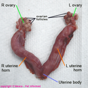
Spaying or desexing is the surgical removal of a female (queen) cat's internal reproductive structures, including her ovaries (site of ova/egg production), Fallopian tubes, uterine horns (the two long tubes of uterus where the foetal kittens develop and grow) and a section of her uterine body (the part of the uterus where the uterine horns merge and become one body).

This is an image of a feline uterus that has been removed by cat spaying surgery.

Part of female reproductive organ that have been removed via spaying

Basically, the parts of the female reproductive tract that get removed are those which are responsible for egg (ova) production, embryo and fetus development and the secretion of the major female hormones (oestrogen and progesterone being the main ones).

Removal of these structures plays a big role in feline population control; feline genetic disease control; the prevention and/or treatment of various medical disorders and female cat behavioral modification (e.g. estrogen is responsible for many female cat behavioral traits that some owners find problematic - e.g. roaming, calling for males - and spaying, by removing the source of female hormones like estrogen, may help to resolve these issues).


Spaying procedure

1. Anesthesia

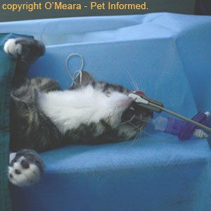
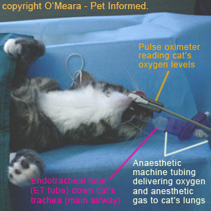
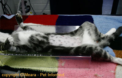
The feline is anesthetized prior to cat spaying surgery.


During a spaying procedure, the cat is placed under anesthetic so that it will not feel pain whilst spay surgery is being performed.

The cat must be anesthetised prior to spay surgery being performed, both so that it will not move whilst the spaying procedure is being performed and also so that it will not experience any pain.

The cat is given a series of injectable sedative and general anesthetic drugs to make it go to sleep (fall unconscious); an endotracheal (ET) tube is placed down its trachea (main airway) to help it to breathe better and to keep its airway free of vomit and other secretions and the cat is maintained under anesthesia by the addition of anesthetic gas vapours to the oxygen that it breathes (the oxygen and anesthetic gas vapors are supplied by an anesthetic machine, which is linked to the cat's endotracheal tube).

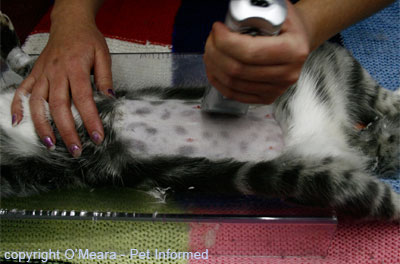
2. Shave belly-to remove the fur at the site of spaying

The fur is clipped from the cat's belly prior to a cat spaying procedure being performed.

The final product - the belly is completely shaved and almost ready for feline desexing surgery to begin.


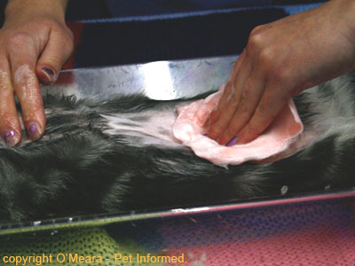
3. Scrub the surgical site

The spay site is scrubbed and cleaned with an antibacterial solution prior to surgery to reduce bacterial contamination of the cat spay site.

Female cat's abdomen (surgical site) being scrubbed with an antiseptic, antibacterial solution (chlorhexidine scrub and alcohol) prior to desexing surgery. This pre-surgical skin preparation reduces the amount of bacterial contamination that is present on the skin prior to the first incision being made.


4. Draping the cat spay site

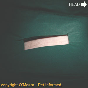
A close-up picture of the cat spaying procedure site.


Cat spay procedure image - A drape is placed around the surgical cat spay site just prior to the first incision being made.


A sterile surgical drape is placed around the surgical site. This drape acts to focus the veterinary surgeon's attention on the spay site. It also acts to cover up the non-surgically-prepared, contaminated regions (e.g. the furred, unclipped regions) located outside of the shaved and prepped site so that the veterinarian can not accidentally touch them and inadvertently contaminate the surgical site. Additionally, the drape also provides a sterile surface for the vet to rest instruments periodically during surgery.


5. Incised the skin

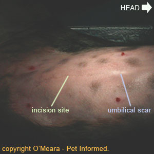
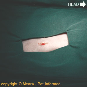
The first incision is made during the cat spaying procedure.

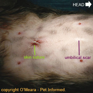
A small incision (usually around 1cm long, but can be up to 3-4 cm long) is made in the cat's skin, approximately 1 inch below the umbilical scar on the abdominal midline.

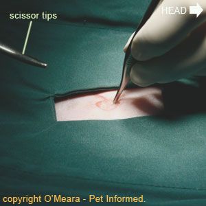
Feline spay image - The veterinary surgeon is removing some of the subcutaneous fat from the incision line region

In this image, the veterinary surgeon is removing some of the fat (termed subcutaneous or SC fat) from the incision line region. The fat is the white, shiny substance in the centre of the incision line. There is generally a lot of fat located between the cat's skin and its abdominal wall muscles. The veterinarian will often cut a small amount of this fat away, allowing easy access to and visualisation of the cat's abdominal wall muscles.


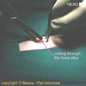
Cat spaying procedure picture - The abdominal wall is incised along its midline, by cutting along the linea alba (white line).

The veterinarian enters the cat's abdominal cavity by cutting through the abdominal wall musculature on the midline of the abdomen. The veterinarian aims to cut along a central line of scar tissue that joins the right and left sides of the animal's abdominal wall musculature. This line of scar tissue is called the linea alba (literally meaning - "white line"). By cutting through scar tissue, rather than the red muscle located either side of the linea alba, the veterinarian reduces the amount of bleeding incurred in entering the cat's abdominal cavity.

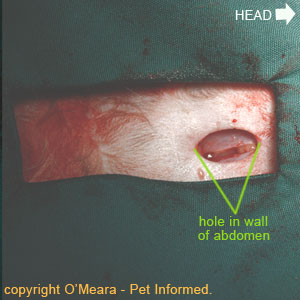
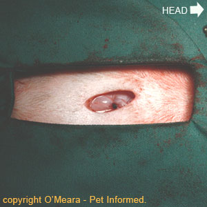
Spaying cats image - This is a close-up image of the incised abdominal wall, showing the hole entering the cat's abdominal cavity.


6. The 1st uterine hone is revealed

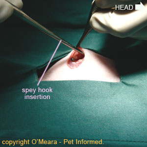
Feline spay procedure - A spay hook is inserted into the cat's abdominal cavity to hook and draw up the first uterine horn.


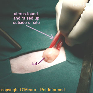
The first feline uterine horn is revealed.

First uterine horn being lifted up and drawn out through the abdominal incision line.


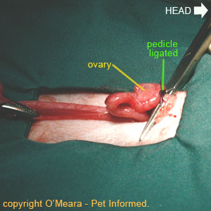
7. The ovarian blood vessels are clamped and ligated

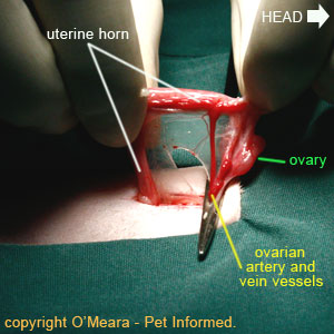
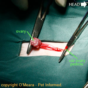
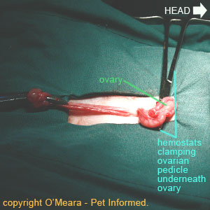
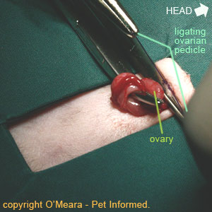
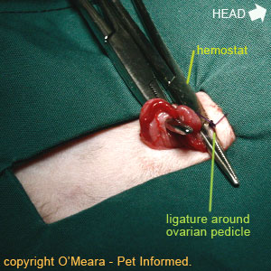
Feline spaying procedure picture - the ovarian pedicle (a term for the blood vessels supplying the ovary) is clamped off using hemostats.

The blood vessels (artery and vein) supplying the cat's ovary are elevated and clamped off using mosquito hemostats (artery forceps). These hemostat clamps crush and traumatize the ovarian blood vessels, causing them to spasm and narrow in diameter, thereby aiding in preventing excessive ovarian pedicle hemorrhage when the ovary is cut off.

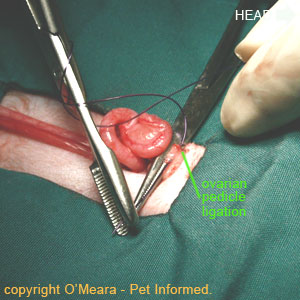
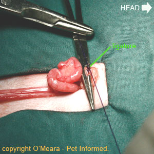
Cat spaying procedure - A suture is placed around the ovarian blood vessels supplying the feline ovary.

A suture (stitch) is placed around the blood vessels supplying the ovary (the general term for the blood vessels - artery and vein - supplying the ovary is the ovarian pedicle). This suture ties off and occludes the ovarian blood vessels supplying the ovary, thereby preventing excessive ovarian pedicle hemorrhage when the ovary is cut off.

During cat spaying, the ovarian pedicle is ligated (tied off with sutures).

Suture (also called a ligature) being placed around the blood vessels supplying the ovary. This suture ties off and occludes the ovarian blood vessels supplying the ovary, thereby preventing excessive ovarian pedicle hemorrhage when the ovary is cut off. Once the ligature has been tied and knotted tightly, the long suture ends are trimmed away leaving only a small knot behind

The ligature has been placed around the ovary pedicle during the cat spaying procedure and the long suture ends have been trimmed away.


8. The ovarian pedicle is cut above the suture

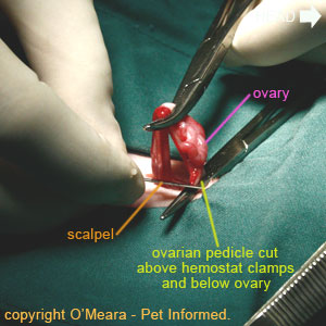
Cat spay image - A scalpel blade is used to cut through the ovarian pedicle (ovarian artery and vein) supplying the ovary.

A scalpel blade is used to cut through the ovarian pedicle (ovarian artery and vein) supplying the ovary. The cut is made above the level of the hemostat clamp and the ovarian pedicle ligature so that the blood vessels (in particular, the ovarian artery) will not bleed when they are incised, but below the ovary such that the ovary will be removed from the ovarian pedicle when the cut is made.

The ovarian pedicle has been cut with the scalpel.

The appearance of the ovarian pedicle after the cut has been made. The hemostats are still in place in this image with the ligature located beneath them (hidden from view in this pic) - the hemostats will be removed, allowing the ovarian pedicle and its ligature to return back inside the abdomen. The ovary, still attached to its uterine horn, is reflected caudally (towards the animal's tail).


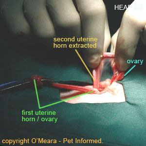
9. Steps 6-8 are repeated for the second uterine horn

Spaying cats - The second uterine horn is lifted up and drawn out through the cat's abdominal spay incision line.

Second uterine horn being lifted up and drawn out through the cat's abdominal spay incision line.

Desexing female cats - The second ovarian pedicle is clamped off above the level of the ovary.

The blood vessels (artery and vein) supplying the cat's second ovary are elevated and clamped off using mosquito hemostats (artery forceps). These hemostat clamps crush and traumatise the ovarian blood vessels, causing them to spasm and narrow in diameter, thereby aiding in preventing excessive ovarian pedicle hemorrhage when the second ovary is cut off.

Feline spay image - the second ovarian pedicle in the process of being ligated (tied off).

Cat spay image - the second ovarian pedicle is ligated (the knot is clearly visible in this image).

As occurred with the first ovarian pedicle, a suture (also called a ligature) is placed around the blood vessels (termed the ovarian pedicle) supplying the second ovary. This suture ties off and occludes the ovarian blood vessels supplying the ovary, thereby preventing excessive ovarian pedicle hemorrhage when the ovary is cut off. Once the ligature has been tied, the long suture
ends are trimmed away, leaving a small knot behind

Following the placement of this ligature, a scalpel is then used to cut through the ovarian pedicle supplying the second ovary. The cut is made above the level of the hemostat and the ovarian ligature (suture) so that the blood vessels (in particular, the ovarian artery) will not bleed when they are incised, but below the ovary such that the ovary will be removed (along with the uterine horn) from the ovarian pedicle when the cut is made.


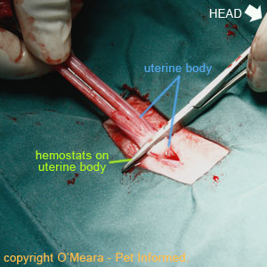
10. The uterine body is revealed and ligated

Spaying cats image - The two uterine horns are pulled caudally and the uterine body is clamped with hemostats.

The two uterine horns are pulled caudally (towards the cat's tail) until the uterine body (the place where the two uterine horns merge and become one uterus body) is revealed and elevated above the level of the skin incision (where it is easily accessible to the surgeon). One or more hemostats are clamped across the uterine body, below the level of the uterine horns and just above the level of the cervix (the cervix is a sphincter-like muscle band located further down the uterine body, which forms a physical barrier between the abdominally-located uterus and the pelvically-located vagina).

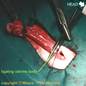
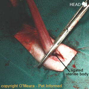
Feline desexing - The uterine body is ligated.

A suture (ligature) is placed around the uterine body. The suture's role is to close off the tunnel leading into the uterus from the outside world. This will prevent bacteria from entering the abdominal cavity, via ascension from the vagina, once the uterus is removed. The uterine body ligature also acts to occlude the uterine blood vessels (the uterine arteries and veins), which run along each side of the uterine body and supply the uterus, thereby stopping them from hemorrhaging once the uterine body has been excised (cut off).

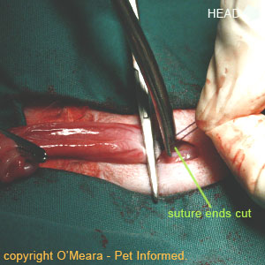
The ligature is now tied and knotted around the feline uterine body. The surgeon is now cutting the long suture ends away from the knot.

The ligature is now tied around the feline uterine body and the surgeon is just cutting the long suture ends away from the knot. Vet surgeons don't like to leave a lot of excess suture material lying around inside an animal's abdomen. Excess suture material can lead to irritation and inflammation occurring inside the abdomen and cause adhesions (where the organs get stuck together by scar tissue) to form between organs.

Spaying procedure pic - The ligated feline uterine body.

The appearance of the ligature once the long suture ends have been cut off the knot.


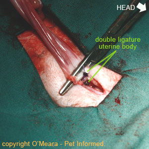
11. A second ligature is placed around the uterine body

Double ligating in spaying procedures. A second ligature is placed around the uterine body for added protection.


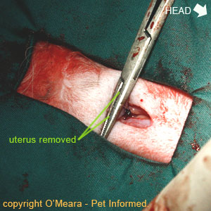
12. The uterine body is cut-off

The uterine body is transected (cut off) above the level of the ligatures.

The uterine body is transected (cut off) above the level of the ligatures. This essentially completes the process of removing the uterus from the female cat. The animal will now no longer be able to reproduce. This is an irreversible surgical procedure.


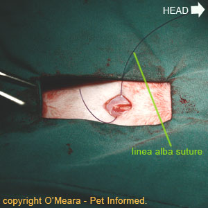
13.The abdominal wall is sutured closed

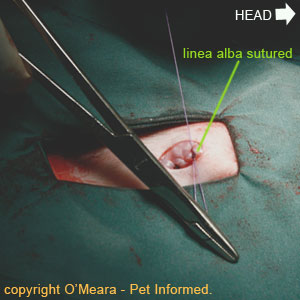
Cat spay image - closing the linea alba and abdominal midline.

Female feline spaying image - closing the linea alba and abdominal midline.

The surgeon uses absorbable suture material to close the hole in the abdominal wall musculature (linea alba). Because the linea alba is essentially a tendon-like, collagenous structure (made of collagen), it has less blood supply than red muscle and, therefore, takes longer to heal than muscle would. To take this slower healing into account, the veterinarian often uses a longer-lasting suture (a suture that is slower to lose its strength and slower to absorb) to close the linea alba. Because this suture absorbs over time, the vet does not have to remove it later on.

Spaying procedure image - The appearance of the linea alba once it has been sutured closed.

The linea alba has been sutured closed.


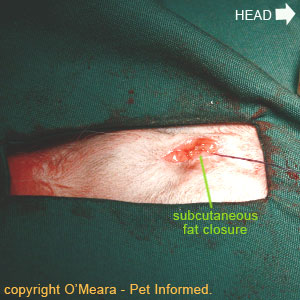
14. The subcut fat layer is sutured close

The subcutaneous fat layer (sub-q or SC layer) is closed to reduce dead space.

The subcutaneous fat layer (also called the SC or sub-q layer) is sutured closed. This layer closure acts to reduce the amount of open space (called 'dead space') located between the abdominal wall and skin layers, thereby reducing the risk of a large, fluid-filled swelling (called a seroma) forming at the surgery site. Basically, whatever space/gap you leave in a surgery site, fluid will pool in - by closing down this open space (dead space), the vet surgeon essentially leaves fewer sites available for inflammatory fluids to pool in.


15. The skin layer is sutured close

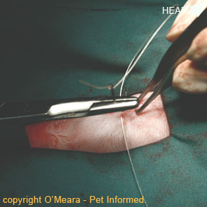
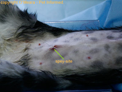
Cat spaying procedure image - The surgeon is closing the skin using non-absorbable skin sutures.


Feline spaying procedure - The skin is closed with non-absorbable skin sutures.

The surgeon is closing the skin using non-absorbable skin sutures. These will need to be removed in 10-14 days.

NOTE - absorbable skin sutures can also be placed. These are called intradermal sutures and they do not need to be removed.


The final result : The newly desexed cat

The newly sterilized female cat.


ps: I will post about cat castration (male cat's) in the later post


ps: A veterinary guide to cat spaying procedure; www.pet-informed-veterinary-advice-online.com

  • Digg
  • Del.icio.us
  • StumbleUpon
  • Reddit
  • RSS
Post Icon

Feline Panleukopenia Virus

Just got to know that one of UMK's stray cat (the one I've posted about rectal palpation) died because of feline panleukopenia.

**********************************************************


" Are you sick?? please tell me..hurmm"

awww soo cute :)


Feline panleukopenia (also called feline infectious enteritis, feline "distemper," and feline ataxia or incoordination) is a highly contagious viral disease of cats characterized by its sudden onset, fever, inappetence (loss of appetite), dehydration, depression, vomiting, decreased numbers of circulating white blood cells (leukopenia), and often a high mortality rate. Intrauterine (within the uterus) infection may result in abortions, stillbirths, early neonatal deaths, and cerebellar hypoplasia (underdevelopment of the cerebellum) manifested by incoordination (ataxia) in kittens beginning at two to three weeks of age.

All members of the cat family (Felidae) are susceptible to infection with feline panleukopenia virus (FPV), as are raccoons, coatimundis, and ringtails, in the family Procyoniclae.


Etiology and transmission

Feline panleukopenia virus is a very small and very stable virus classified in the parvovirus group. The genetic material of the virus is composed of a single"strand of DNA. The virus is highly resistant to most disinfectants ether, chloroform, acid, alcohol, and heat (56'C, or 132.8'F, for thirty minutes)but is susceptible to Clorox bleach. Replication (reproduction) of the virus in the host occurs in cells that are themselves actively reproducing. Incubation period is between 5-10 days. Primary site of infection are in the rapidly dividing cells which includes cerebellum, lymphoid tissue and intestine. It may reduce white blood cells number.


Feline leukopenia virus caused by Parvoviridae family


FPV is a severe, highly contagious disease that is oftentimes fatal. Feline panleukopenia occurs worldwide, but is rarely seen as a clinical entity due to the effectiveness of vaccination in preventing the disease. Young, unvaccinated kittens present most commonly with this disease. Unvaccinated feral cat colonies and other wild felids also serve as reservoirs of infection for the domestic cat population.

FPV exposure and infection can occur in several ways. The major route of transmission is direct contact between a susceptible host and an infected animal or its secretions. The virus is shed in all body secretions of infected animals for up to six weeks. Once introduced into the environment, the virus is very hardy and can persist for years.

Treatment of fomites (inantimate objects) and other contaminated materials for ten minutes with bleach, 4% formaldehyde or 1% gluteraldehyde is necessary to inactivate the virus. Fomites, including contaminated instruments, cages and bedding, are also an important route of viral exposure. Mechanical transmission of FPV via arthropod vectors is probable as well. Lastly, this virus also can cross the placenta to infect the fetuses in utero.


Pathogenesis

The virus usually enters orally, with infection occurring primarily in the lymphoid tissues of the oropharynx (tonsillar area) and intestine. Within twenty-four hours of infection, virus is present in the blood, which distributes it throughout the body. Within two days of infection, nearly every body tissue contains significant amounts of virus. As circulating antibodies appear, the amount of virus present gradually decreases. Small quantities of virus may persist for up to one year in certain tissues, but the strong immune response of the host usually neutralizes the virus as it is shed, so that most persistently infected kittens are not infectious.

The most severely damaged tissues in the infected newborn cat are those undergoing rapid cell division -the thymus (lymph organ in the chest) and the cerebellum (rear of the brain). Cells of the small intestine, which have a slow turnover rate in neonates (newborns), are not damaged, although the virus is present within them. In older kittens, the development of the disease also depends on the reproductive activity of the various tissues within the body. Lymphoid tissues, bone marrow, and the surface cells of the intestine are the most severely affected.

Clinical signs

  • Fever
  • Diarrhea
  • Vomitting
  • Rough, dry skin (coat)
  • Loss of appetite
  • Ataxia
  • Swollen abdomen

Diagnosis

  • Basis of history
  • Clinical signs
  • Blood cell count
  • Fecal test
  • Virus isolation
  • Necropsy examination

Treatment

Panleukopenia normally has a high mortality rate, but with diligent effort and good nursing care this can often be reduced. The main objective is to keep the affected animal alive and in reasonably good health until the natural defenses take over (i.e., the appearance of antibodies and an increase in number of circulating white blood cells). Antibodies usually appear about three to four days after the first signs of illness; two to three days later, the sharp "rebound" in white blood cell number can be expected to occur.

Thus, if the cat can be supported for five to seven days after onset of the disease, the chances of recovery usually are good. Veterinary supportive care is aimed at the vomiting, diarrhea, and dehydration, which may dangerously upset fluid and electrolyte balance, and at preventing secondary bacterial infections. Secondary viral respiratory infections are common complications of panleukopenia. The FPV infection may act to trigger a latent respiratory virus, such as feline viral rhinotracbeitis virus or feline calicivirus. Simultaneous FPV and respiratory virus infections usually produce a more severe illness than if either virus alone had infected the animal.


Prevention

There are several excellent vaccines available to immunize cats against panleukopenia. These vaccines are highly effective and produce long-lasting immunity. Because panleukopenia is an entirely preventable disease, one cannot overemphasize the importance of proper immunization.Immunization should be initiated by the veterinarian when kittens are eight to ten weeks of age. A second vaccination should be given four weeks later.


FPV's Vaccines

In areas where the prevalence of infection is high, and for maximal protection, a third vaccination may be indicated at sixteen weeks of age. if a kitten is twelve weeks of age or older at the time of initial vaccination with a modified live virus vaccine, a booster vaccination need not be given until it is at least one year of age. Immunity produced by FP vaccines is long-lasting, perhaps for life.

Revaccinations every year would not seem to be necessary from a scientific standpoint, but the vaccines are licensed for only 3 years' protection.Immunity acquired from the queen via colostrum (initial breast milk) must be considered when establishing a routine vaccination program. interference by maternally acquired (passive) immunity is the most common cause of vaccine failure. There exists a direct correlation between the FPV antibody level of the queen at the time of birth and the duration of passive immunity in the kitten. This passive immunity, if of sufficient strength, will not only protect the kitten against virulent FPV but will also react with the vaccine virus and interfere with immunization.

Vaccination must be performed after kittens have lost most or all of their maternally derived immunity.The use of FPV antiserum (clear blood liquid containing antibody) to immunize cats passively is indicated if an unvaccinated animal has been exposed to the virus or is likely to be exposed before vaccine induced immune responses can develop. Antiserum is also indicated for colostrum-cleprived or orphaned kittens. The routine use of antiserum in unexposed kittens is not recommended, however; instead, kittens should be vaccinated during their first visit to the veterinarian's office, and revaccinated as indicated.


Sources: Max's house; FPV, Catworld; www.cat-world.com.au, feline panleukopenia, Veterinary clinical pathology clerkship program; FPV

  • Digg
  • Del.icio.us
  • StumbleUpon
  • Reddit
  • RSS