Powered by Blogger.
RSS

Welcome

I created this blog as an instrument of what I have encountered in the world of veterinary medicine as a proud vet student. Comments and suggestions are welcome here at;

sweet_daffodil90@yahoo.co.uk

Regards,
Aina Meducci 2012

Disclaimer

The following blog posts is not genuinely from my research but through readings and citation from trusted website. I do not own any of the copyright and therefore you may use it at your own risk

SINCE I AM NOT A VETERINARIAN YET, THEREFORE I CAN'T CONSULT ANY MEDICAL ADVICE TO YOU AND YOUR PETS! EXTREMELY IMPORTANT!.

Happy reading!
Post Icon

Teething (Veterinary dental) Part 1

I was thrill to found out indeed there are Veterinary dental collage situated somewhere in the US. In Malaysia, veterinary field is still a small sector and so we have developed less veterinary specialties but I believe we are progressively moving towards a promising veterinary future, one day insyaAllah.

******************************************************************
Nomenclature of specific teeth

The incisors will be referred to as: (right or left) (maxillary or mandibular) first, second, or third incisors numbered from the midline.

The central incisor is always 01 and the following incisors are 02 and 03.The canines are always 04.The premolars are 05 to 08 and the last premolar is always 08.The first molar is always 09 and the following molars are 10 and 11 (The Tridan system)


The Tridan system in cat


The existence of the conventional anatomical names of teeth as well as the various tooth numbering systems is recognized. The correct anatomical names of teeth are (right or left), (maxillary or mandibular), (first, second, third or fourth), (incisor, canine, premolar, molar), as applicable, written out in full or abbreviated.

The modified Triadan system is presently considered to be the tooth numbering system of choice in veterinary dentistry; gaps are left in the numbering sequence where there are missing teeth (for example, the first premolar encountered in the feline left maxilla is numbered 206, not 205. The two lower right premolars are 407 and 408, not 405 and 406).


Surfaces of teeth and directions of mouth

Vestibular is the correct term referring to the surface of the tooth facing the vestibule or lips; buccal and labial are acceptable alternatives.

Lingual: The surface of a mandibular or maxillary tooth facing the tongue is the lingual surface. Palatal can also be used when referring to the lingual surface of maxillary teeth.

Mesial and distal are terms applicable to tooth surfaces. The mesial surface of the first incisor is next to the median plane; on other teeth it is directed toward the first incisor. The distal surface is opposite from the mesial surface.

Rostral refers to a structure closer to, or a direction toward the most forward structure of the head. Caudal refers to a structure closer to, or a direction toward the tail.


Generations of teeth in diophyodont species

Diophyodont: A Diphyodont is an animal with two successive sets of teeth, first the "deciduous" set and later the "permanent" set. Most mammals are Diphyodonts. Diphyodonts contrast with Polyphyodonts, such as many fish, whose teeth are constantly replaced.


The deciduous dentition period (milk teeth) is that period during which only deciduous teeth are present. The mixed dentition period is that period during which both deciduous and permanent teeth are present. The permanent dentition period is that period during which only permanent teeth are present.


Deciduous teeth


Retained teeth?

The primary tooth should always be shed as the permanent tooth cuts through the gum. But sometimes the root of the primary tooth does not dissolve and the tooth remains firmly held in the jaw. The permanent tooth then “glances off” the retained primary tooth and erupts through the gum at an improper angle.


All retained teeth should be extracted as soon as the condition is recognized. If the extractions are performed early, the abnormally positioned adult tooth usually moves over to fill the void and assumes a more correct position. The removal of retained deciduous teeth is an inexpensive, simple way to prevent major problems from developing in the adult dentition.

If retained primary teeth are allowed to remain in the mouth, the teeth become crowded, rotated, or tilted at abnormal angles.

This will result in;
  • early onset and increased severity of gum disease
  • damage to the soft tissues of the mouth, due to sharp teeth penetrating unprotected gum and mouth tissues
  • pain, in the joints of the jaw as well as in the gums, lips, and teeth
  • excessive wear, when abnormally aligned teeth grind against other teeth and weaken them


Jaws, salivary glands and lymph nodes

All mammals have two maxillas (or maxillae) and two mandibles.


Mandibula

All animals have two mandibles, not one -- removing one entire mandible therefore is a mandibulectomy not a hemimandibulectomy


Corpus mandibulae (body of mandible)

The part that carries the teeth - often incorrectly referred to as horizontal ramus


Pars incisive (incision part)

The part that carries the incisors


Pars molaris (Molar part)

The part that carries the premolars and molars premolar-molar part would probably have been more accurate


Margo alveolaris (margin of alveolar)

Alveolar crest


Margo ventralis

Ventral margin


Canalis Mandibulae (canal of mandibulae)

Contains only the neurovascular bundle often incorrectly referred to as the medullary cavity of the mandible


Foramina mentalia (mental foramen)

Rostral, middle or caudal mental foramina in the dog and cat


Ramus mandibulae (Ramus of mandible)

The part that carries the 3 processes often incorrectly referred to as the vertical ramus


Synchondrosis intermandibularis (mandibular symphysis)

Collum mandibulae (Neck of mandible)


Incisive bones

In domestic animals, the correct name for the paired bones that carry the maxillary incisors, located rostral to the maxillary bones, is the incisive bones, not the premaxilla.


Mandibular salivary glands and nodes

Domestic animals have a mandibular gland (or mandibular salivary gland) and a mandibular lymph node. The term "submandibular," as used in humans, is incorrect due the difference in topography of these structures.


Fauces

The fauces are defined as the lateral walls of the oropharynx that are located medial to the palatoglossal folds. The areas lateral to the palatoglossal fold, commonly involved in feline stomatitis, are not the fauces.


Hard palate

The midline of the hard palate is not a symphysis but is formed by the interincisive suture, the median palatine suture of the palatine processes of the maxillary bones, and the median suture of the palatine bones.


Dental fracture classification


To be continue..

Sources: AVDC nomenclature & Virginia Veterinary Dentistry


  • Digg
  • Del.icio.us
  • StumbleUpon
  • Reddit
  • RSS
Post Icon

Feline digestive system


I have been asked by my lecturer during oral vet med examination few weeks ago. I remember her asking about cat digestive system. Her question was

" How long the food retain in the digestive tract until it'll be secreted out?"

My answer were simple: "24 hours..maybe?.." (actually i wasnt sure if that was the correct answer)

Then she gave me a long sharp look that almost kills my gaze and says;

"WRONG.."

Here we go..

**********************************************************

Feline digestive system


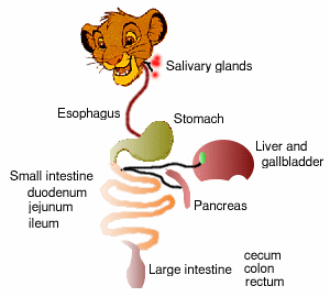
Cats are true carnivores. Their digestive system plays two important roles: it breaks down food into nutrients that the cat's body can readily absorb, and it also acts as a protective barrier against harmful bacteria and other pathogens (disease causing agents) that the cat may ingest.

The digestive system includes


Mouth


Teeth


Esophagus and stomach


Small and large intestine


Pancreas


Liver


Gross anatomy of cat's digestive system


1. Diaphragm

The muscle which expands and contracts in order to allow the cat to breathe. It separates the thoracic and abdominal cavities

2. Gallbladder

The gallbladder stores bile until it is needed by the small intestine in order to digest fats. The gallbladder is not essential to the process of digestion. It is possible for bile to be delivered directly from the liver to the small intestine. Bile digests and absorbs fat and helps to dispose of waste from the cat's body.

3. Liver

The liver produces many substances in order to digest food. It manufactures bile, which is then stored in the gall bladder. Bile contains acids as well as cholesterol. The liver also stores and releases a compound called glycogen, which is a carbohydrate that can be changed into sugar should the sugar level of the cat fall.


4. Pancreas

The pancreas secretes three compounds. The first compound is a digestive enzyme that helps break down fats and proteins. The second compound is insulin, which regulates the sugar level in the cat's body. The third is a bicorbonate ion that nutralizes stomach acid


5. Peritoneum (not part of GIT)

The peritoneum is the thin membrane that covers the abdominal digestive organs of the cat.

6. Small intestine

The small intestine carries out the final stages in digestion. It is lined with small hairs and blood vessels inside the intestine carry away nutrients to other parts of the cat's body. The small intestine has three sections: the duodenum, the jejunum, and the ileum. The duodenum is the section where the process of absorption begins. The jejunum is the largest portion of the small intestine and it is here where most of the absorption takes place. The ileum is the final section of the intestine and it is where the absorption process ends.


7. Spleen (not part of digestive)

The spleen produces red blood cells and regulates the supply of red blood cells. It also functions as a storage area for blood.


8. Stomach

The stomach stores food while it goes through the initial phases of digestion.


The arrangment of feline digestive


How does the digestion begins?


Humans and Cats are similar in their digestive process in the sense that when food enters the mouth large amounts of saliva are secreted. The saliva will lubricate, dissolve, and chemically break down the food. As a cat takes food into its mouth, it chews it into small pieces. The tongue will assist moving the food to its oral pharynx, the hollow structure at the back of its oral cavity. From there, the food is swallowed, passing into the esophagus--a tube that travels down through the animal's chest and opens into its stomach.


Within the stomach, the ingested food mixes with potent acids and enzymes that are produced by the stomach lining. Feline gastric juices are more powerful than those of a human; they are, in fact, strong enough to soften bone. Cats can swallow large chunks of prey creatures (rodents and birds) and any parts such as feathers, hair and bones that are not quickly broken down in the stomach may be regurgitated.


Once the food passes through the stomach, it then moves through the pylorus, a sphincter, or ringlike muscle, that constricts or relaxes as needed to control flow into the first section of the small intestine (duodenum). This is where most of the digestion takes place. There the food mixes with bile, a bitter yellowish liquid secreted by the liver, the largest organ in a cat, and with a fluid produced by the pancreas. These substances and their enzymes play a key role in neutralizing the harsh stomach acids and breaking down proteins, fats and carbohydrates so that they can be absorbed through the intestinal lining (mucosa) and into the cat's bloodstream. The liver also purifies toxins that have entered the system and recycles dead red blood cells.


From the small intestine, the food passes into the large intestine, which absorbs additional nutrients and also a substantial amount of the water remaining in the ingested food. The large intestine serves as a storage area for the solid material that is left over after the digestive process. This material--the feces--is eventually excreted from the cat's body, through its rectum and anus.


This process normally takes about 20 hours in an adult cat.


Get the answer?


Here something I found on the web about feline nutrition requirements

For adult maintenance

Unless otherwise listed, all values are minimum requirements:

Protein... 26%
Fat ...... 9%
Calcium.... 0.6%
Phosphorus... 0.5%
Potassium... 0.6%
Sodium..... 0.2%
Chloride.... 0.3%
Magnesium... 0.04%
Iron... 80 mg/kg
Copper... 5 mg/kg
Manganese.... 7.5 mg/kg
Zinc....... 75 mg/kg (maximum 2000 mg/kg)
Iodine..... 0.35 mg/kg
Selenium.... 0.1 mg/kg
Vitamin A... 5000 IU/kg (maximum 750,000 IU/kg)
Vitamin D... 500 IU/kg (maximum 10,000 IU/kg)
Vitamin E... 30 IU/kg
Thiamine... 5 mg/kg
Riboflavin... 4 mg/kg
Pantothenic Acid... 5 mg/kg
Niacin... 60 mg/kg
Pyridoxine... 4 mg/kg
Folic Acid....0.8 mg/kg
Vitamin B12...0.022 mg/kg
Choline..... 2400 mg/kg
Taurine... 0.1%

For Growing Kittens, Pregnant and Lactating Queens

The majority of nutrient minimums are the same except for the items listed. The maximum for those listed does not change.

Protein...30%
Calcium 1%
Phosphorus 0.8%
Magnesium... 0.08%
Copper... 5-15 mg/kg
Vitamin A... 9000 IU/kg
Vitamin D... 750 IU/kg

I'll post about feline nutrition later.

Sources: How the cat's digestion works; examiner.com, Dietary requirements in cat petplace.com, Digestive system of the cat, Washington state university


  • Digg
  • Del.icio.us
  • StumbleUpon
  • Reddit
  • RSS
Post Icon

Basic surgical instruments

Hello again dear readers! Today I want to share briefly about basic surgical instruments. I realized that I haven't posted anything regarding surgery yet and I thought this is going to be a good start.

Note: This webpage I found is basically show the instruments which are used in veterinary medicine field.

***************************************************************

Surgical instruments exist in vast numbers and varieties due to many types of operation such as laparotomy, rumenotomy, gastrotomy and many more.

The following are examples of the basic instruments that should learned by every beginning surgeon.


1. Scissors

Classification by types of points


borsh.GIF (12665 bytes)

All types of scissors can have blunt or sharp blades (A: Sharp:Sharp, B: Blunt:Blunt)


Blade Shapes

All types can have either straight or curved blades.


1. Mayo and Matsembaum


Mayo and Metzenbaum Scissors

Mayo scissors (B) are used for cutting heavy fascia and sutures.
Metzenbaum scissors (A) are more delicate than Mayo scissors.

Metzenbaum scissors are used to cut delicate tissues.Metzenbaum scissors have a longer handle to blade ratio.


2. Forceps

Consist of two tines held together at one end with a spring device that holds the tines open. Forceps can be either tissue or dressing forceps.

Dressing forceps have smooth or smoothly serrated tips.Tissue forceps have teeth to grip tissue. Many forceps bear the name of the originator of the design, such as Adson tissue forceps.


Rat tooth forceps


Adsons


Interdigitating teeth hold tissue without slipping.
Used to hold skin/dense tissue.


Adson tissue forceps


Rat tooth

Small serrated teeth on edge of tips.The Adsons tissue forceps has delicate serrated tips designed for light, careful handling of tissue.


Allis tissue forceps


Allis

Interdigitating short teeth to grasp and hold bowel or tissue.Slightly traumatic, use to hold intestine, fascia and skin.


Babcock forceps


Babcock

More delicate that Allis, less directly traumatic.Broad, flared ends with smooth tips.Used to atraumatically hold viscera (bowel and bladder)


Sponge forceps


Sponge Forceps

Sponge forceps can be straight or curved.Sponge forceps can have smooth or serrated jaws.
Used to atraumatically hold viscera (bowel and bladder).


Hemostatic forceps

They are used to clamp and hold blood vessels.


Hemostat Tip Shape

Hemostatic forceps and hemostats may be curved or straight.


Kelly and mosquitoes forceps


Kelly and Mosquito

Both are transversely serrated.
Mosquito hemostats (A) are more delicate than Kelly hemostatic forceps (B).

Kelly and Mosquito Tips

Mosquito hemostats (A) have a smaller, finer tip.


Carmalt forceps

Carmalt

Heavier than Kelly.Preferred for clamping of ovarian pedicals during an ovariohysterectomy surgery because the serrations run longitudinally.


Doyen intestinal forceps

Doyen Forceps

Doyen intestinal forceps are non-crushing intestinal occluding forceps with longitudinal serrations.Used to temporarily occlude lumen of bowel.


Payr Pylorus forceps

Crushing Forceps

Payr pylorus clamp is a crushing intestinal instrument.
Used to occlude the end of bowel to be resected.


Needle holder

Hinged (locking) instrument used to hold the needle while suturing tissue.

Mayo Hager (most common use)

Align Center

Mayo-Hegar Needle Holders

Heavy, with mildly tapered jaws.
No cutting blades.


Olsen Hager

Olsen-Hager Needle Holders

Includes both needle holding jaw and scissors blades.The disadvantage to having blades within the needle holder is the suture material may be accidentally cut


Scalpel Handle and blades

Scissor Points

No3 and No4 scalpel handle


Blade Shapes

Blades #10, 11, 12, 15 fit the #3 handle.
Blades #22, #23 fit the #4 handle and are commonly used for large animals


Disposable Scalpel


Towel clamps

Backhaus Towel CLamp

Locking forceps with curved, pointed tips.
Towel clamps secure drapes to a patient's skin. They may also be used to hold tissue.


  • Digg
  • Del.icio.us
  • StumbleUpon
  • Reddit
  • RSS
Post Icon

White muscle disease (WMD)

Assalam, Hello people!! Dr Meducci is back! Geez, I really miss blogging!! I've been stressed up for the whole month for final examination and it was AWEEEEso.... erm actually, no target to be shoot this time, I just hope the result will be as good as the previous semester.


Damn! My surgery and pathology T.T

*****************************************************************

Today i would like to share regarding White Muscle Disease. The reason why I choose WMD because this topic was often discussed in my class. Let's get a review of it


08.jpg (19333 bytes)

White muscle disease


White muscle disease (WMD) is a degenerative muscle disease found in all large animals. Commonly it is found in grazing lamb or sheep It is caused by a deficiency of selenium and/or vitamin E. Generally, it is not known which. Selenium (Se) deficiency is associated with selenium deficient soils and the inadequate uptake of selenium by forages grown on these soils.

So it is all about the NUTRITIONAL problem


Cause of disease

Pasture, hay, grain, and other supplements can be analyzed to determine the amount of selenium to be added to supplemental feeds. Grazing sheep usually consume adequate amounts of vitamin E. Fresh legumes and pasture are good sources of vitamin E whereas silage, oil seeds, root crops, cereal grains, and dry hays tend to be poor sources of vitamin E. Prolonged storage of feedstuffs results in a degradation of Vitamin E content.

In addition to white muscle disease, selenium and vitamin E deficiencies can produce symptoms of ill thrift and reproductive losses. They can cause poor rate of growth or ill thrift in young lambs throughout the growing period. Selenium and vitamin E also play key roles in the animal's normal immune response.


White Muscle Disease

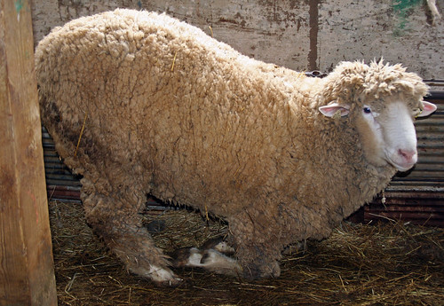
Lameness of lamb


While the leg muscles are generally affected first, any muscle area including cardiac muscle may be affected. Chronic or borderline deficiencies may result in lung edema resulting in increased pneumonia problems. It is quite common for the disease to show up after forced exercise, vaccination procedures, sorting, weaning, fitting or being chased by dogs or children.


Normal heart (left) vs WMD heart (right)


Clinical signs

All breeds of sheep and goats are suceptible to WMD, and the condition may develop under extensive or intensive management systems. WMD is most commonly found in newborns or fast growing animals. Kids are believed to be more susceptible than lambs, possibly because they have a higher requirement for selenium. The disease can affect both the skeletal and cardiac muscles.

When the skeletal muscles are affected, symptoms vary from mild stiffness to obvious pain upon walking, to an inability to stand. Lambs/kids may tremble in pain when held in a standing position. A stiff gait and hunched appearance are common. Affected lambs/kids may remain bright and have normal appetites, but eventually they become too weak to nurse. When the problem occurs in newborns, they are born weak and unable to rise. Sudden exercise may trigger the condition in older lambs and kids.

When the disease affects the heart, the animal shows signs similar to pneumonia, including difficult breathing, a frothy nasal discharge (may be blood stained), and fever. The heart and respiratory rates are elevated and often irregular. Skeletal and cardiac muscle disease may occur concurrently.

The congenital type of white muscle disease may result in sudden death within 2-3 days of birth, usually with involvement of the myocardium. The delayed type is associated with cardiac or skeletal muscle involvement and may be precipitated by vigorous exercise. Affected animals may move stiffly with an arched back and frequently become recumbent.

If the condition is severe enough to prevent nursing, either from dysfunction of the muscles of the legs or the tongue, death may result from starvation. Sometimes, there is profuse diarrhea. In chronic cases, there may be relaxation of the shoulder girdle and splaying of the toes. In progressive cardiac failure, dyspnea results. Signs vary with dietary selenium status; in some areas, general unthriftiness may be the only sign associated with selenium deficiency.


Hunched appearance is affected sheep

Stiff gait


Diagnosis

Selenium deficiency can be confirmed by measuring selenium levels in whole blood or tissues. A diseased animal will have less than 0.04 ppm of selenium in its blood. Breeding ewes require more selenium, and their blood levels should be over 0.5 ppm. At necropsy, the muscles of affected animals appear paler than normal and may show distinct longitudinal striations or a pronounced chalky appearance due to abnormal calcium deposition.


Paler flesh of dead affected sheep


Histolopath finding: necrosis of muscle


Review pathology: how does it occur???

Deficiency in vitamin E and/or Selenium → peroxidation of membrane lipids (sometimes exacerbated by stress) → Ca 2+ enters cytoplasm and mitochondria, damaging respiratory mechanisms → cell death → symmetrical necrosis of the most active skeletal muscle (like postural muscles)


Treatment

Lambs and calves may be given sodium selenite and vitamin E in sterile emulsion, SC or IM, at 1 mg selenium and 50 mg (68 IU) of vitamin E per 18 kg (40 lb) body wt. This may be repeated after 2 wk, but no more than 4 doses should be given. Larger dosages are sometimes advocated, but caution is advised because they approach the toxic level. In practice, several products are available for use with designated animal species. When simple vitamin E deficiency is apparent, dietary supplementation with α-tocopherol or substances rich in vitamin E should be instituted. Minimum dosages have not been established.

When the causative diet contains substances antagonistic to vitamin E, such as unprotected, polyunsaturated fats, these must be removed or stabilized by addition of an appropriate antioxidant. Dry concentrates of vitamins A and D may substitute for cod-liver oil, thus removing a potential source of oxidative damage.


Prevention

To prevent white muscle disease within 4 wk after birth, ewes are given 5 mg and cows 15 mg of selenium, PO or SC, usually as sodium selenite 4 wk before expected parturition. To prevent the delayed type, lambs are given 0.5 mg and calves 5 mg of selenium at 2-4 wk of age and twice more at monthly intervals. A selenium and vitamin E mixture is advocated in some areas. Other procedures for selenium supplementation include administration of intraruminal selenium pellets, use of selenium-fortified salt or mineral mixtures, SC implantation of selenium pellets, or soil application of selenium at 4 g/acre (10 g/hectare) in fertilizer.

Adding selenium to feed for breeding animals or their young is useful in areas of known deficiency. The recommended supplemental level is 0.3 ppm selenium, calculated on the basis of total dry-matter intake. It is added as sodium selenite, which contains 45.65% selenium. Because of the minute quantities involved and the toxicity of excess intake, premixing and thorough subsequent mixing is necessary. In some countries, caution in the use of selenium is indicated.

Sources: Barbados Blackbelly Sheep Association, University of Maryland;white disease in sheep and goat, Merck Veterinary Manual., WMD; purdue.edu


  • Digg
  • Del.icio.us
  • StumbleUpon
  • Reddit
  • RSS